INSIDE EMBRYO · IVF & Fertility Basics
Start with the cells.
Understand the foundations.
Get to know eggs, sperm, reproductive anatomy and the processes that connect them. A clear starting point for patients, students and curious readers.

LOOK. READ. UNDERSTAND.
Explore the foundations.
Follow ten illustrated topics at your own pace. The image introduces the idea; the explanation helps you connect it to the bigger picture.
This image could not load. Use the full-size image link above to try opening it directly.
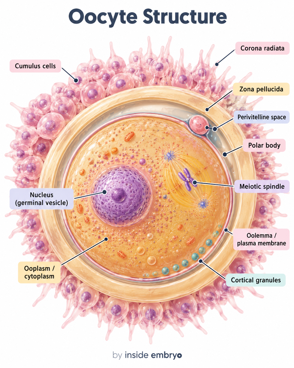
Oocyte Structure
The oocyte is the female reproductive cell. This illustration introduces structures such as the zona pellucida, ooplasm, polar body, cortical granules and the surrounding cumulus cells.
This image could not load. Use the full-size image link above to try opening it directly.
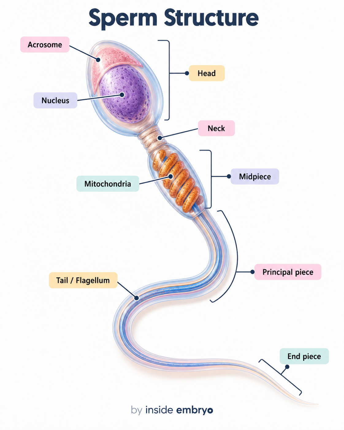
Sperm Structure
A sperm cell contains specialised regions for fertilisation and movement. Explore the acrosome, nucleus, neck, mitochondrial midpiece and different regions of the flagellum.
This image could not load. Use the full-size image link above to try opening it directly.
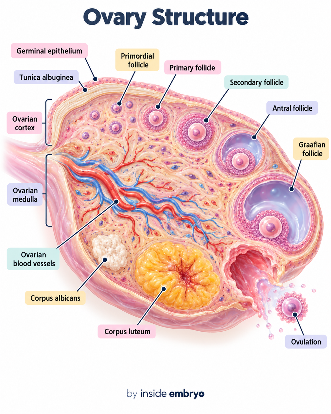
Ovary Structure
The ovaries contain follicles at different developmental stages. The diagram shows the ovarian cortex and medulla, developing follicles, blood vessels, corpus luteum and ovulation.
This image could not load. Use the full-size image link above to try opening it directly.
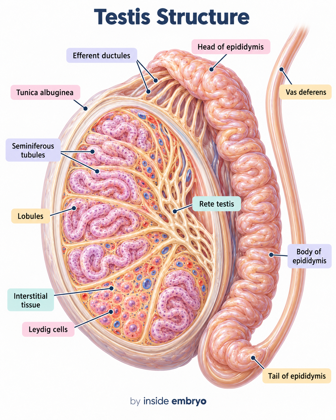
Testis Structure
The testis contains seminiferous tubules where sperm production occurs. The illustration also introduces the rete testis, epididymis, Leydig cells and vas deferens.
This image could not load. Use the full-size image link above to try opening it directly.
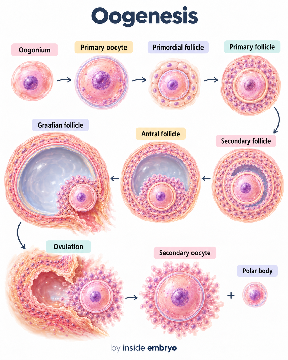
Oogenesis
Oogenesis describes the development of the female germ cell. The illustration follows the relationship between the developing oocyte, follicular growth, ovulation and the secondary oocyte.
This image could not load. Use the full-size image link above to try opening it directly.
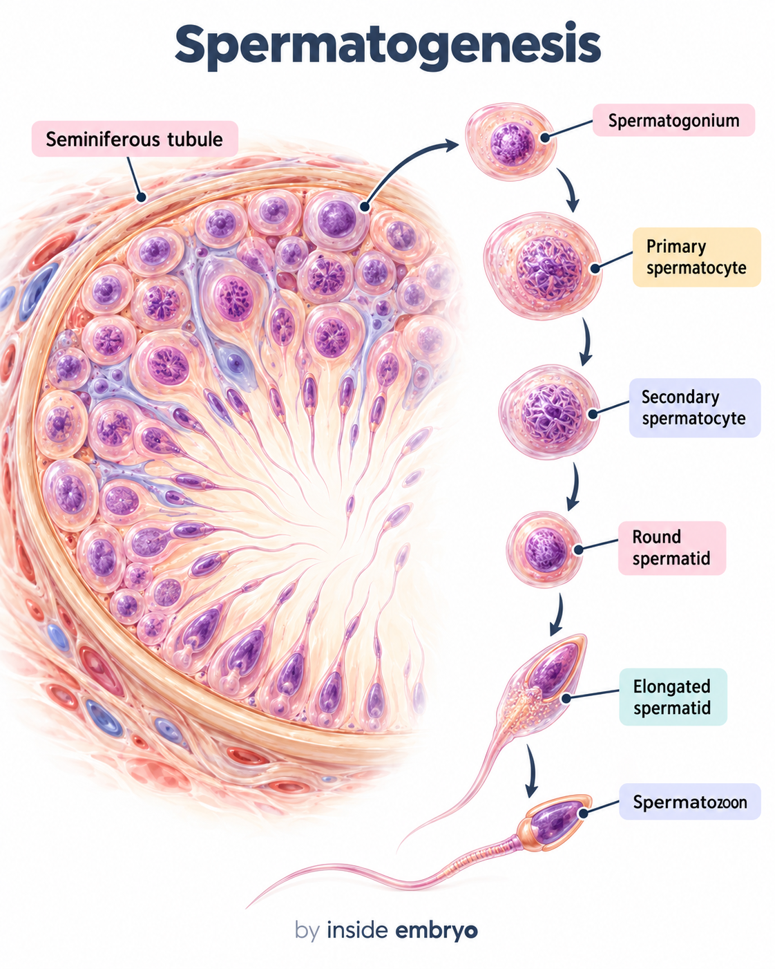
Spermatogenesis
Spermatogenesis produces spermatozoa from germ cells within the seminiferous tubules. Sperm then undergo further functional maturation during passage through the epididymis.
This image could not load. Use the full-size image link above to try opening it directly.
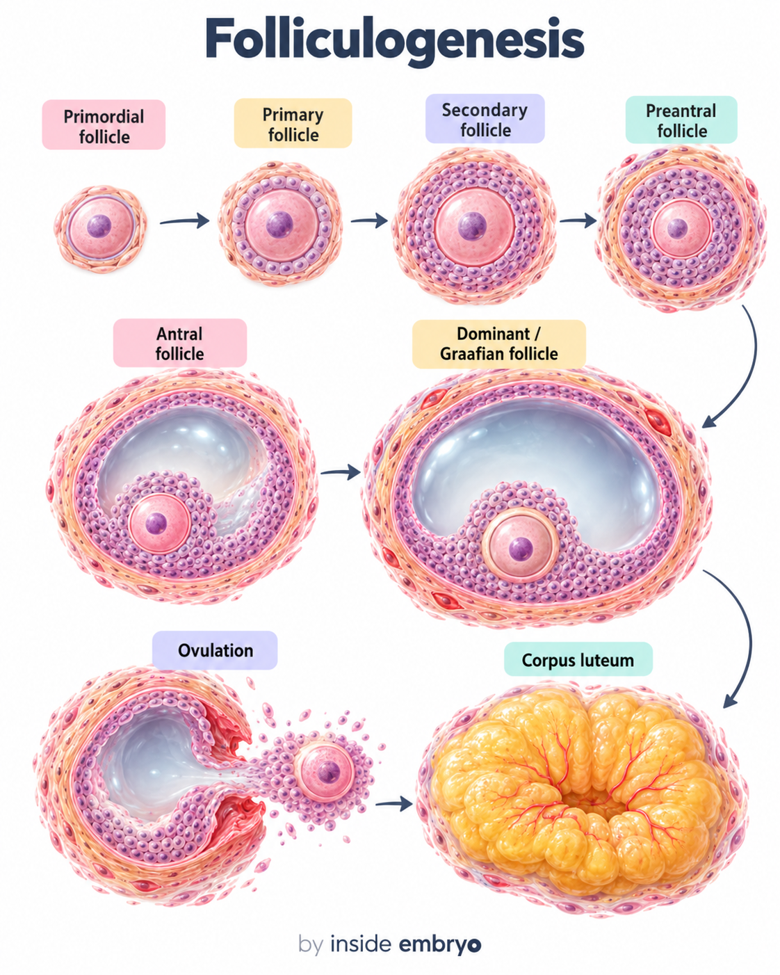
Folliculogenesis
Folliculogenesis describes the development of ovarian follicles through successive stages. In a typical ovulatory cycle, a selected dominant follicle may ovulate and form a corpus luteum; many other follicles undergo atresia.
This image could not load. Use the full-size image link above to try opening it directly.
Menstrual Cycle
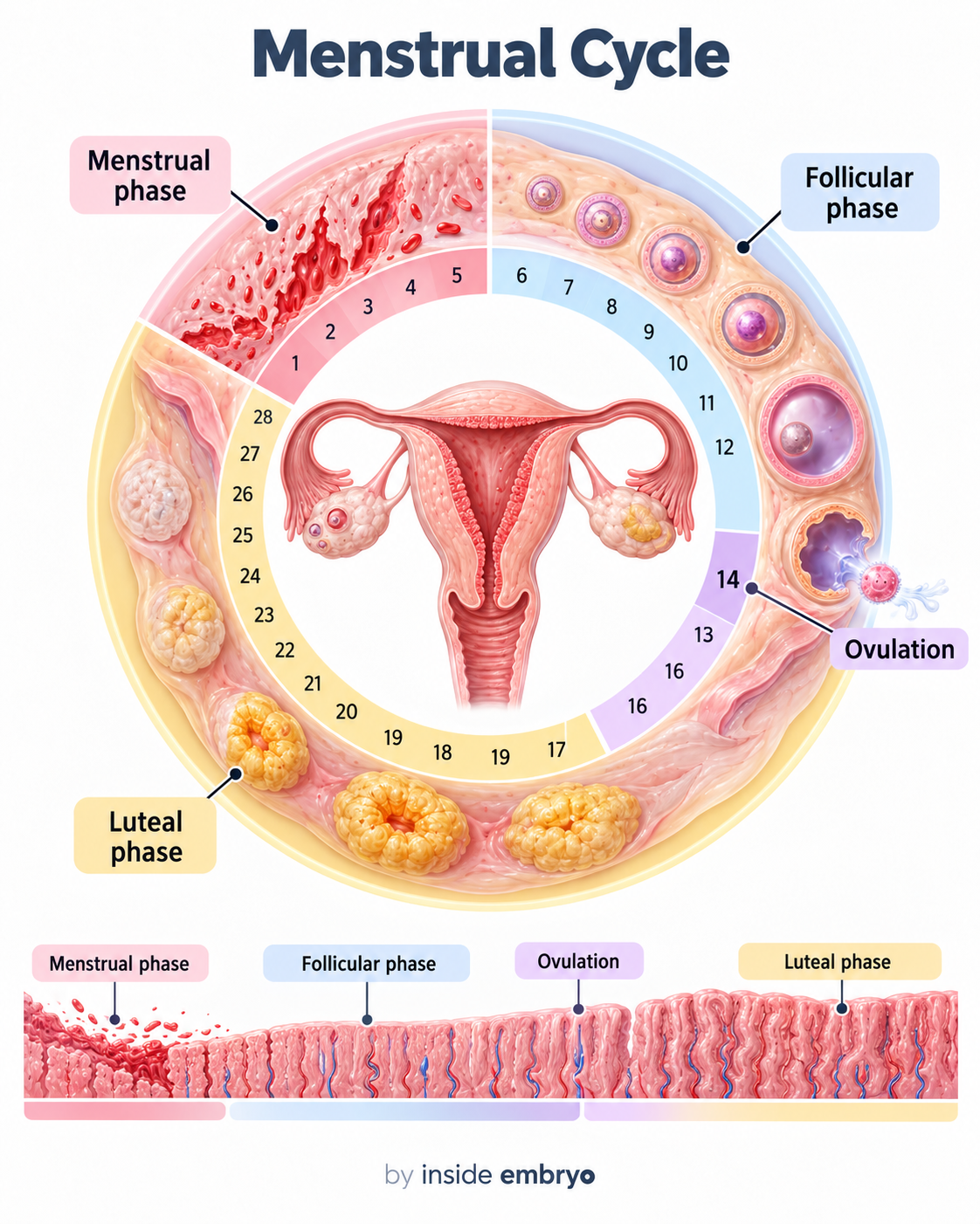
The menstrual cycle coordinates changes in the ovaries and endometrium. The major stages include menstruation, the follicular phase, ovulation and the luteal phase.
This image could not load. Use the full-size image link above to try opening it directly.
Ovulation Process
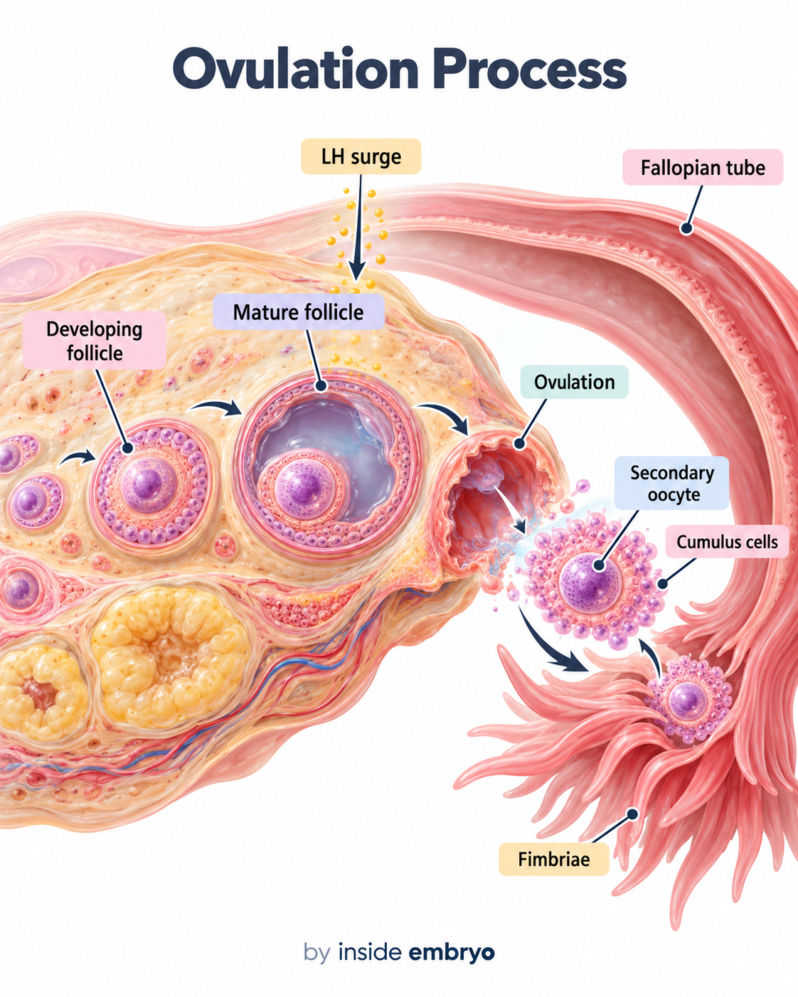
Ovulation is the release of the secondary oocyte from the dominant follicle. After release, the cumulus–oocyte complex is positioned near the fimbrial end of the fallopian tube.
This image could not load. Use the full-size image link above to try opening it directly.
Blastocyst Structure
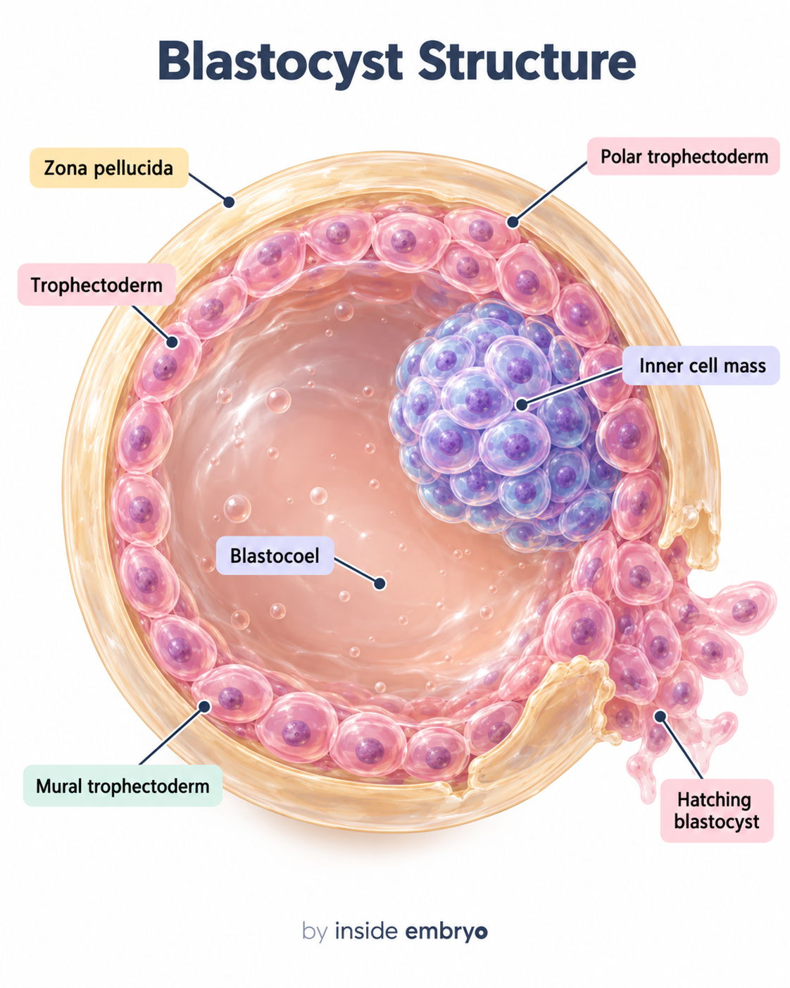
A blastocyst contains a fluid-filled blastocoel, an inner cell mass and an outer trophectoderm layer. Expansion is followed by thinning and eventual hatching from the zona pellucida.
Swipe across the pictures. On a computer, use the arrows or focus the carousel and press ← / →.
CONTINUE YOUR READING
The latest in IVF & Fertility Basics.
A little more detail, one article at a time.
The latest published article will load here on the website.
A LITTLE CLARITY GOES A LONG WAY
You may have heard…
Let’s look at the facts.
Five common misunderstandings about ivf & fertility basics. Explore the facts beside each one.
01 / IVF & Fertility Basics
IVF and implantation are the same event.
IVF involves fertilisation outside the body. Implantation is a later process in the uterine lining; transferring an embryo does not confirm that implantation has happened.
02 / IVF & Fertility Basics
ICSI is the best fertilisation method for everyone.
ICSI can be useful for specific indications, including some sperm-related problems. It is not automatically better for every patient or every cycle.
03 / IVF & Fertility Basics
Every follicle seen on a scan will provide an egg.
The scan and the collection tell the team different things. Some follicles may not yield an egg, so the final egg count can differ from the scan count.
04 / IVF & Fertility Basics
Embryo freezing is used only when a cycle has failed.
Freezing can be part of a planned treatment strategy or preserve suitable additional embryos for later use. The reason depends on your individual plan.
05 / IVF & Fertility Basics
A positive pregnancy test means no follow-up is needed.
A positive test is an important milestone. Your clinic still arranges follow-up to assess how the pregnancy is progressing and discuss the next steps.
EXPLORE MORE IN THIS CATEGORY
Keep learning about IVF & Fertility Basics.
Take the next question a little further. Browse the articles below, with the newest reads first.
Articles from this category load here. You can also use the archive link below.
CONNECT THE IDEAS
Where would you like to go next?
Patient Education
Turn the basic science into questions for your care team.
Explore Patient Education →IVF Journey
Follow the overall patient pathway from assessment to follow-up.
Explore IVF Journey →Andrology
Explore sperm biology, semen assessment and preparation.
Explore Andrology →TAKE A QUESTION WITH YOU
From reading
to a clearer conversation.
Which structure or process are we discussing?
How does it relate to the treatment being considered?
Which term would I like explained more clearly?
A little understanding makes the next question easier.
You can return to these illustrations whenever a term feels unfamiliar. Build your knowledge one idea at a time.
Sources and notes about this page
- HFEA · IVF explained
- HFEA · ICSI explained
- NHS · Egg collection
- HFEA · Embryo freezing
- NHS · Pregnancy testing and follow-up
- ESHRE · Good practice in IVF laboratories (2015)
- OpenStax · Embryonic development
Illustrations explain concepts and are not diagnostic images, grading tools or laboratory protocols. Some diagrams simplify structures or timing. Your care team interprets findings from your own treatment.
By Manoj Kumar K, Embryologist · Medical disclaimer · Back to top ↑