
INSIDE EMBRYO · Embryology
Observe the detail.
Understand the science.
Explore oocyte morphology, fertilisation, embryo assessment and genetic testing. Connect each observation with what it can tell us—and what it cannot.

LOOK. READ. UNDERSTAND.
A closer look, one image at a time.
Ten illustrated topics connect laboratory observations with the science of early development. Open an image at full size when you want to inspect its labels.
This image could not load. Use the full-size image link above to try opening it directly.
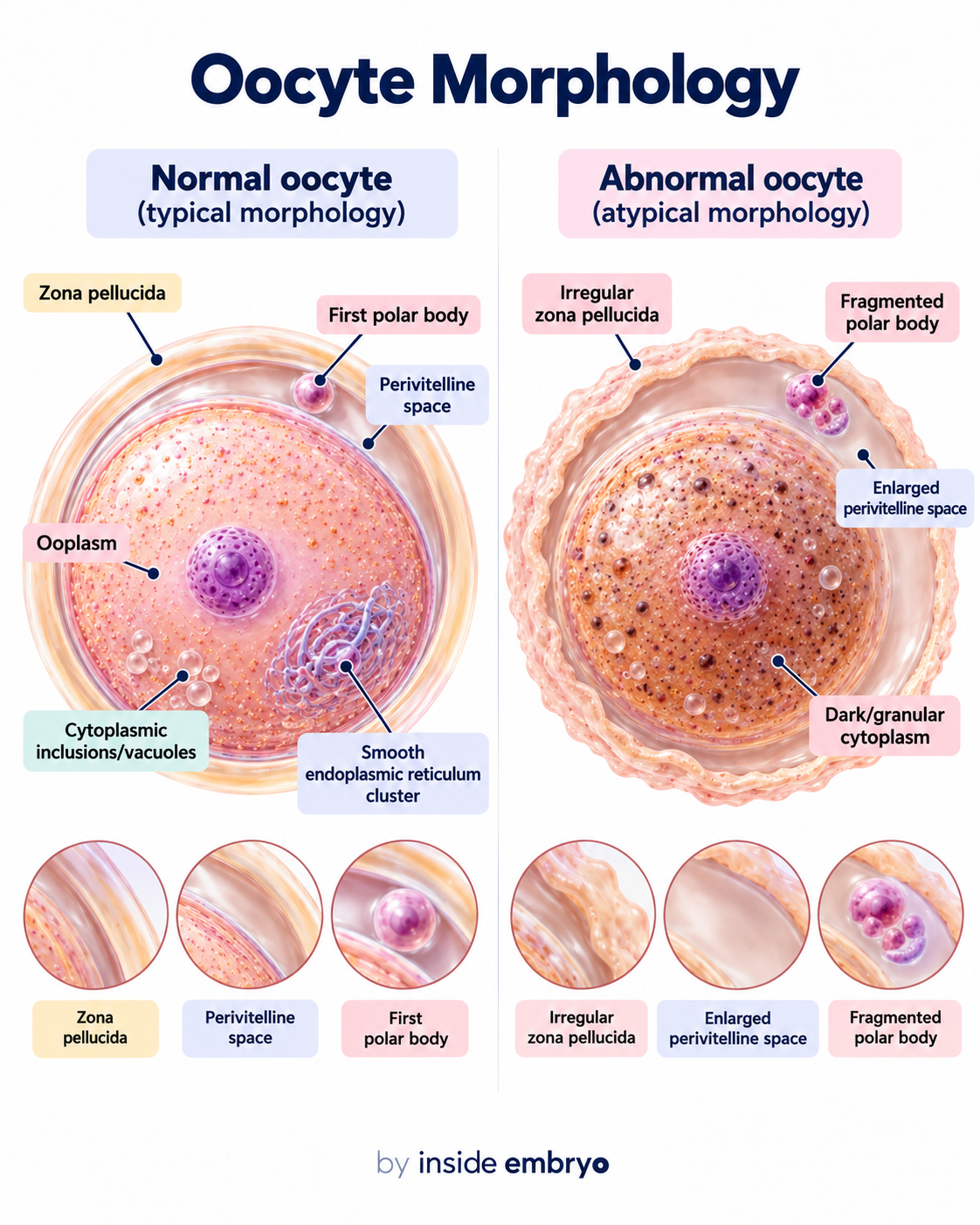
Oocyte Morphology
Oocyte morphology is assessed by observing features such as the ooplasm, zona pellucida, perivitelline space and polar body. Cytoplasmic granularity, vacuoles and other morphological variations may also be recorded.
This image could not load. Use the full-size image link above to try opening it directly.
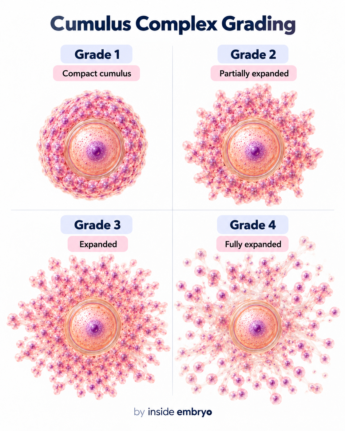
Cumulus Complex Assessment
The cumulus–oocyte complex contains the oocyte surrounded by cumulus and corona cells. The degree of cumulus expansion can change as the follicle and oocyte progress through maturation.
This image could not load. Use the full-size image link above to try opening it directly.
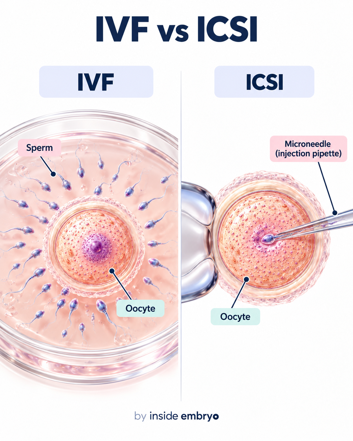
IVF vs ICSI
Conventional IVF and ICSI are laboratory methods used to achieve fertilisation. In IVF, prepared sperm and oocytes are cultured together. In ICSI, a single selected sperm is introduced directly into a mature oocyte.
This image could not load. Use the full-size image link above to try opening it directly.
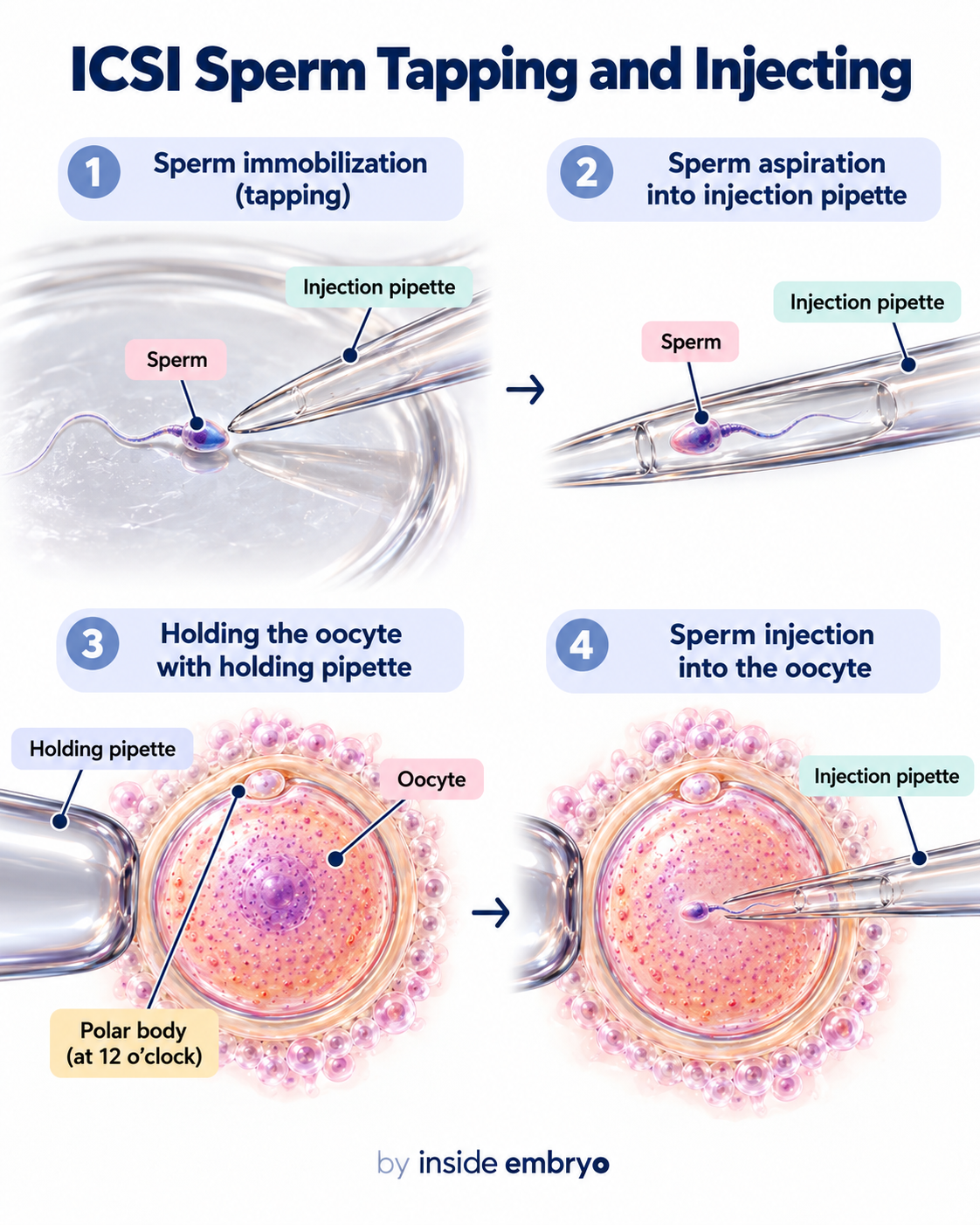
ICSI: Sperm Immobilisation & Injection
ICSI is performed using micromanipulation equipment. A selected sperm is immobilised and aspirated into an injection pipette while the mature oocyte is stabilised using a holding pipette before sperm introduction.
This image could not load. Use the full-size image link above to try opening it directly.
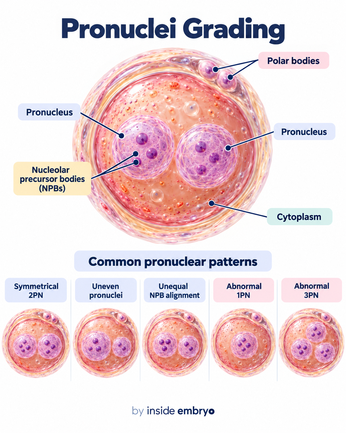
Pronuclear Assessment
Following fertilisation, the zygote can be assessed for the number and appearance of pronuclei and polar bodies. Two pronuclei are the classical morphological finding associated with normal fertilisation.
This image could not load. Use the full-size image link above to try opening it directly.
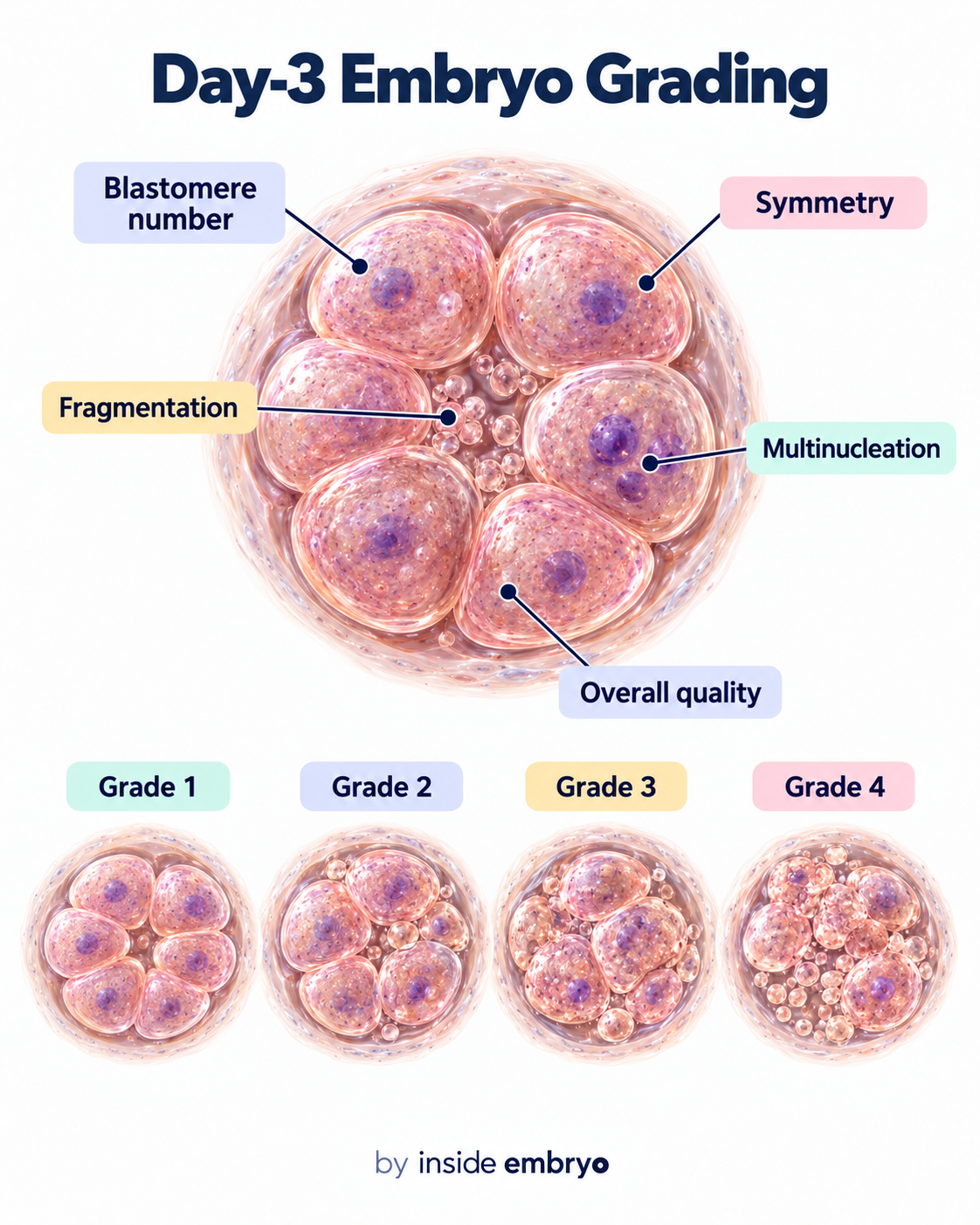
Day-3 Embryo Assessment
Cleavage-stage embryos are assessed by observing features such as blastomere number, blastomere symmetry, fragmentation and multinucleation.
This image could not load. Use the full-size image link above to try opening it directly.
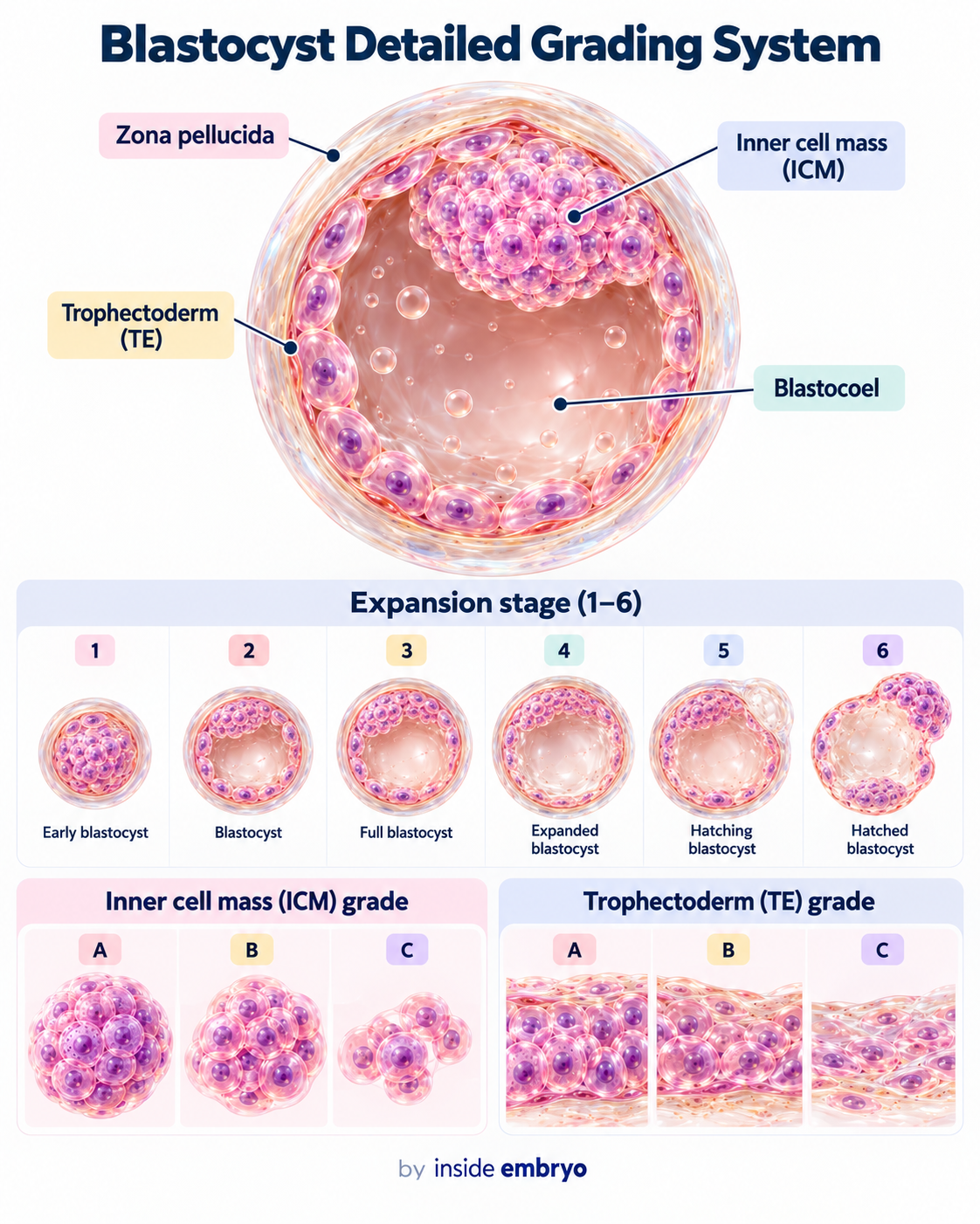
Blastocyst Grading
Blastocyst morphology is commonly described using three components: the degree of expansion, the appearance of the inner cell mass and the appearance of the trophectoderm.
This image could not load. Use the full-size image link above to try opening it directly.
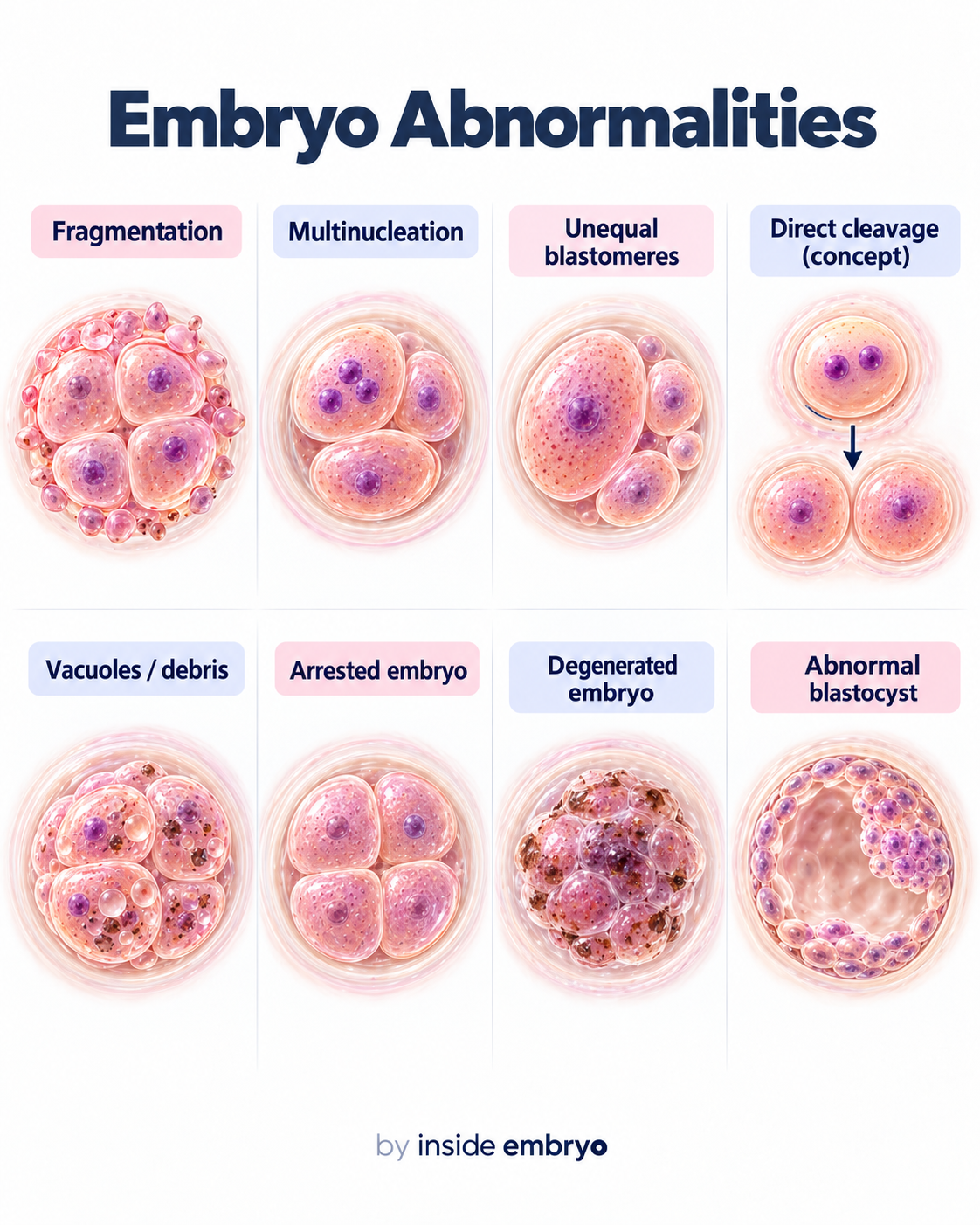
Embryo Morphological Abnormalities
Embryos may show morphological findings such as excessive fragmentation, multinucleation, unequal blastomeres, vacuolisation, degeneration or developmental arrest.
This image could not load. Use the full-size image link above to try opening it directly.
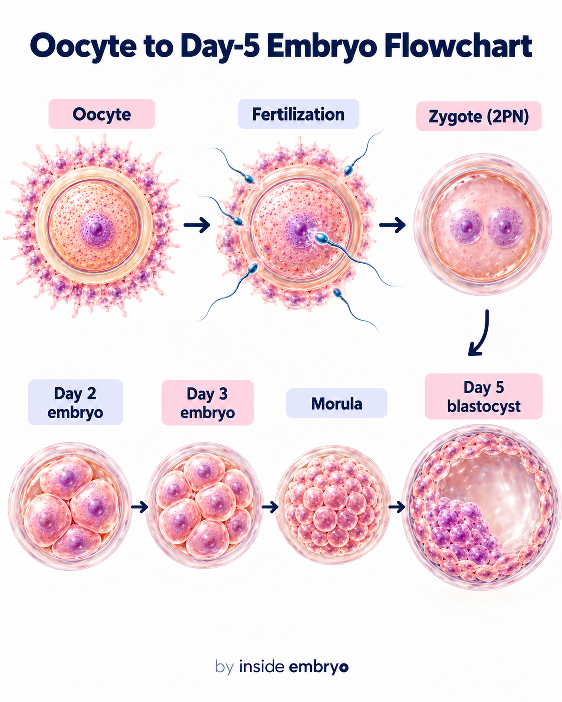
Oocyte to Day-5 Blastocyst
After fertilisation, embryos that continue developing pass through cleavage divisions, compaction and morula formation before becoming blastocysts. Some reach this stage on day 5, others later, and some stop developing.
This image could not load. Use the full-size image link above to try opening it directly.
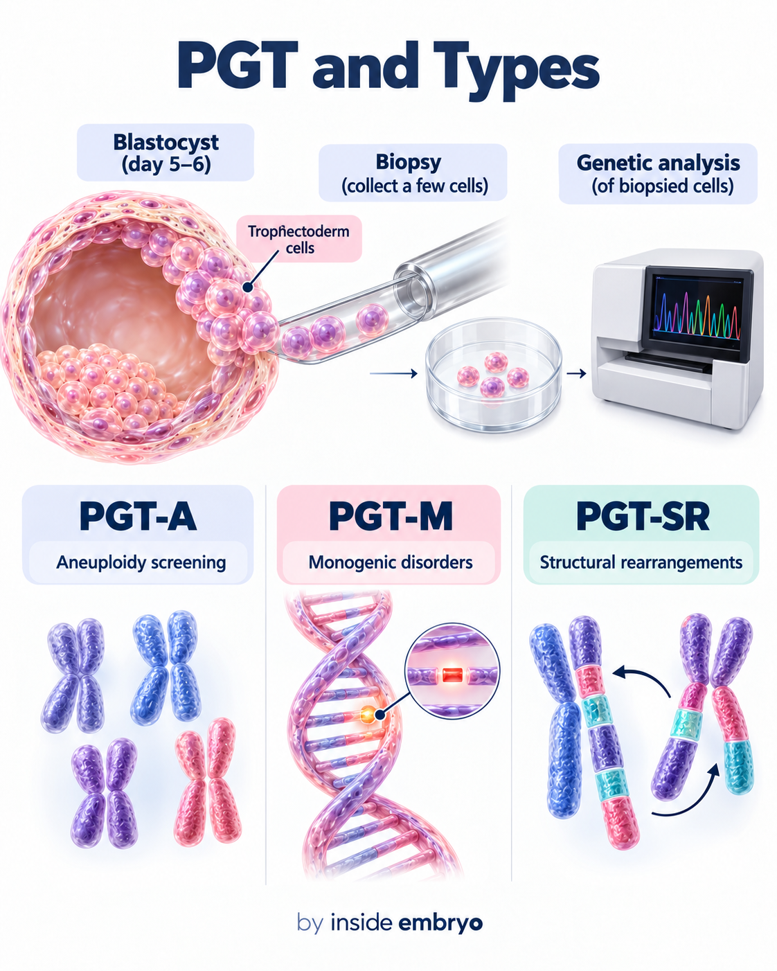
PGT & Its Main Types
Preimplantation genetic testing involves genetic analysis of cells obtained from an embryo biopsy. Current terminology includes PGT-A for aneuploidy, PGT-M for monogenic conditions and PGT-SR for structural chromosomal rearrangements.
Swipe across the pictures. On a computer, use the arrows or focus the carousel and press ← / →.
CONTINUE YOUR READING
The latest in Embryology.
A little more detail, one article at a time.
The latest published article will load here on the website.
A LITTLE CLARITY GOES A LONG WAY
You may have heard…
Let’s look at the facts.
Five common misunderstandings about embryology. Explore the facts beside each one.
01 / Embryology
Embryology is only about IVF procedures.
Embryology studies early development, including cell division, organisation and differentiation. IVF is one setting in which parts of this science are applied.
02 / Embryology
An unfertilised oocyte and a zygote are the same stage.
An oocyte is an egg cell. A zygote is the single-cell stage after fertilisation. The terms describe different points in the reproductive process.
03 / Embryology
Early cleavage makes an embryo double in overall size each time.
During early cleavage, the existing material is divided into smaller cells. More cells do not mean the total volume doubles with every division.
04 / Embryology
An embryo grade is its chromosome result.
Morphology describes appearance. Chromosome testing examines genetic material from sampled cells; these are different types of information.
05 / Embryology
One photograph shows the complete story of development.
A still image captures one moment. Developmental assessments also consider the stage and timing of observations, rather than treating one picture as a complete history.
EXPLORE MORE IN THIS CATEGORY
Keep learning about Embryology.
Take the next question a little further. Browse the articles below, with the newest reads first.
Articles from this category load here. You can also use the archive link below.
CONNECT THE IDEAS
Where would you like to go next?
Embryo Development
Follow the changes from fertilisation through foetal development.
Explore Embryo Development →IVF Laboratory
See how the laboratory supports handling, culture and assessment.
Explore IVF Laboratory →Andrology
Explore the sperm-related science alongside oocyte and embryo biology.
Explore Andrology →TAKE A QUESTION WITH YOU
From reading
to a clearer conversation.
Which stage is being observed, and when?
Which features are described by this assessment?
What cannot be concluded from appearance alone?
Keep asking what the observation means.
A developmental stage, an observed feature and a predicted outcome are different things. Use these explanations as a starting point for deeper reading and supervised learning.
Sources and notes about this page
- OpenStax · Embryonic development
- NHS · Fertilisation in the laboratory
- HFEA · What PGT-A examines
- ESHRE · Good practice in IVF laboratories (2015)
Illustrations explain concepts and are not diagnostic images, grading tools or laboratory protocols. Some diagrams simplify structures or timing. Your care team interprets findings from your own treatment.
By Manoj Kumar K, Embryologist · Medical disclaimer · Back to top ↑