Manoj Kumar K, Embryologist
Inside Embryo | Scientific and Medical Education
Introduction
Cellular biology is the study of cells—the smallest units that perform the fundamental processes associated with life. It examines how cells are organised, obtain and use energy, exchange substances, communicate, reproduce, specialise and die. In the human body, cellular behaviour underlies development, tissue function, healing, ageing and disease.
An introduction to cellular biology is valuable because cells connect molecular events to visible biological outcomes. DNA changes can alter proteins; hormones act through cellular receptors; infections use cellular machinery; and many diseases develop when signalling, metabolism, division or cell-death pathways are disturbed.
The connection to reproductive science is direct. Oocytes and spermatozoa are specialised cells, fertilisation is a cellular interaction, and early embryo development depends on cell cycles, membranes, organelles and gene regulation. This article provides an educational framework, not a diagnostic guide or laboratory protocol.
Learning Objectives
- Define cellular biology and the central ideas of modern cell theory.
- Distinguish prokaryotic cells from eukaryotic cells.
- Relate major human-cell structures to their functions.
- Explain membrane transport, gene expression, metabolism and signalling.
- Outline the cell cycle, differentiation and regulated cell death.
- Recognise limitations of cell morphology and laboratory observations.
- Apply basic cell-biology concepts responsibly to reproductive and medical science.
Key Takeaways
- Cells are the basic structural and functional units of living organisms.
- Human cells are eukaryotic and contain specialised compartments called organelles.
- The plasma membrane is a dynamic, selectively permeable boundary.
- Cell function depends on coordinated gene expression, metabolism, transport and signalling.
- Organelles operate as an interacting network rather than isolated parts.
- The cell cycle is controlled by regulatory proteins and checkpoints.
- Cell appearance alone rarely proves function, viability or developmental competence.
- Cellular biology underpins disease mechanisms, embryology and laboratory science.
Table of Contents
Definition and Overview
Cellular biology, or cell biology, studies the structure, molecular organisation and behaviour of cells. It overlaps with biochemistry, molecular biology, genetics, physiology, developmental biology and pathology. Its central question is how molecules become organised into systems that maintain internal order and respond to their environment.
Modern cell theory states that living organisms are composed of cells, that the cell is a basic unit of biological organisation, and that new cells arise from pre-existing cells. Modern research also shows that cells continuously exchange signals, metabolites and mechanical forces with neighbouring cells and the extracellular matrix.[1,2]
Human cells are eukaryotic: most genetic material is enclosed within a nucleus, and many functions occur in membrane-bound compartments. Different cell types share a basic plan but vary greatly in size, shape, organelle content, lifespan and gene-expression pattern.
Important Terminology
| Term | Clear meaning | Practical relevance |
| Cell | The smallest organised unit that performs core biological functions. | Links molecular events with tissue and organ function. |
| Organelle | A specialised cellular structure or compartment. | Separates and coordinates cellular processes. |
| Plasma membrane | The lipid-and-protein boundary surrounding a cell. | Controls exchange, sensing, adhesion and communication. |
| Cytosol | The aqueous internal fluid outside membrane-bound organelles. | Site of many metabolic and signalling reactions. |
| Cytoplasm | The cytosol plus organelles outside the nucleus. | Describes most of the cell interior. |
| Genome | The complete genetic material of a cell or organism. | Provides inherited information used through regulated gene expression. |
| Gene expression | Use of DNA information to produce functional RNA or protein. | Allows cells with similar DNA to perform different functions. |
| Metabolism | The network of chemical reactions within a cell. | Supplies energy and cellular building materials. |
| Homeostasis | Regulation of internal conditions within a functional range. | Supports stable function during environmental change. |
| Signal transduction | Conversion of a signal into a cellular response. | Links receptors to gene activity, metabolism, movement or division. |
| Cell cycle | The regulated sequence of growth, DNA replication and division. | Produces new cells while protecting genome integrity. |
| Apoptosis | A controlled form of programmed cell death. | Removes unnecessary or damaged cells. |
Classification and Major Cell Types
| Category | Examples | Key feature |
| Prokaryotic cells | Bacteria and archaea | No membrane-bound nucleus; generally smaller and structurally simpler. |
| Eukaryotic cells | Animals, plants, fungi and many unicellular organisms | Nucleus and multiple specialised cellular compartments. |
| Somatic human cells | Most body cells | Support tissue structure, metabolism, communication and repair. |
| Germ-line cells | Cells that give rise to oocytes or spermatozoa | Transmit genetic information to the next generation. |
| Stem and progenitor cells | Self-renewing or developmentally restricted cell populations | Maintain tissues and generate differentiated descendants. |
| Terminally differentiated cells | Highly specialised cells such as many neurons or muscle cells | Perform specialised functions and may have limited division capacity. |
Scientific and Biological Background
Chemical organisation of cells
Cells consist mainly of water, ions and carbon-based molecules. Proteins provide structure, catalyse reactions and support transport and signalling. Lipids form membranes and store energy. Carbohydrates contribute to energy supply and recognition. DNA and RNA store, transmit and use genetic information.[1,2]
The plasma membrane
The plasma membrane is a fluid phospholipid bilayer containing proteins, cholesterol and carbohydrate-containing molecules. Its components can move, allowing the membrane to change shape, assemble receptors and form vesicles.[3,4]
Movement across the membrane depends on molecular size, polarity, charge, gradients and transport proteins. Small non-polar molecules diffuse more readily than ions or most polar molecules. Channels, carriers, pumps and vesicles regulate other substances.
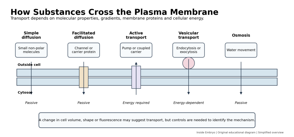
Figure 2. Simplified overview of passive, active and vesicular transport across the plasma membrane.
Cellular compartments and organelles
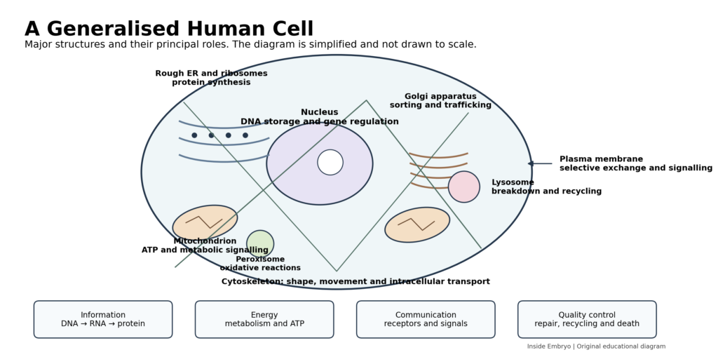
The nucleus stores most cellular DNA and regulates access to genes. The nucleolus supports ribosomal RNA production, while ribosomes translate messenger RNA into protein.
Rough endoplasmic reticulum supports synthesis and processing of many secreted and membrane proteins. Smooth ER participates in lipid synthesis, detoxification and calcium handling. The Golgi apparatus modifies and sorts proteins and lipids.
Mitochondria generate much cellular adenosine triphosphate (ATP) and also influence metabolism, calcium and apoptosis. Lysosomes degrade macromolecules, and peroxisomes perform specialised oxidative reactions. Organelles exchange material through vesicles and direct membrane contact sites.[5]
Cytoskeleton, adhesion and the extracellular environment
Actin filaments, microtubules and intermediate filaments form the cytoskeleton. They support shape, movement, intracellular transport and chromosome segregation. Cell junctions and adhesion molecules connect cells to one another and to the extracellular matrix.
Genetic information and protein production
Gene expression begins when DNA is transcribed into RNA. Protein-coding messenger RNA is processed and translated by ribosomes. Newly made proteins may remain in the cytosol or enter the ER–Golgi pathway. Regulation at several stages allows cells with similar genomes to develop distinct identities.
Energy, metabolism and cellular quality control
ATP couples energy-releasing reactions to transport, movement and biosynthesis. Glycolysis occurs in the cytosol, while mitochondria support oxidative metabolism. Metabolic activity changes with cell type, nutrients, oxygen and developmental state.
DNA repair, protein-folding systems, proteasomal degradation, lysosomes and autophagy help maintain quality. Autophagy recycles selected cellular material, but its biological effect depends on context.
Cell communication and signalling
Cells detect hormones, growth factors, neurotransmitters, matrix signals, nutrients and oxygen. A ligand binds a surface or intracellular receptor, and signal-transduction pathways relay information through protein interactions, phosphorylation, second messengers or gene-expression changes.[6]
The same signal may cause different outcomes in different cells because receptor abundance, developmental state and interacting pathways vary.
Cell cycle, differentiation and death
The proliferative cell cycle includes G1, S, G2 and M phases. DNA is copied during S phase; chromosomes separate during mitosis; and cytokinesis divides the cell. Cyclins and cyclin-dependent kinases regulate transitions, while checkpoints delay progression if DNA is damaged or chromosome attachment is incomplete.[7]
Cells may enter quiescence, differentiate, become senescent or die. Apoptosis uses regulated protease cascades to dismantle cells and supports development and tissue homeostasis. Other forms of cell death have different mechanisms and consequences.[8]
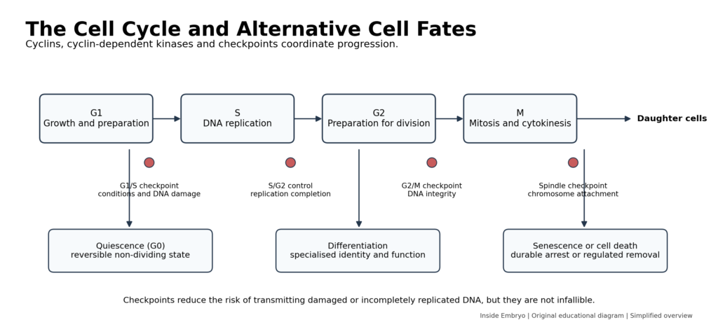
Figure 3. The principal cell-cycle phases, checkpoints and alternative cellular fates.
Process, Mechanism and Cellular Workflow
1. Receive information: Receptors and sensors detect signals, nutrients and physical conditions.
2. Integrate the signal: Intracellular pathways combine new information with the cell’s current state.
3. Regulate gene activity: Transcriptional and epigenetic mechanisms alter RNA and protein production.
4. Build and distribute molecules: Ribosomes, organelles, vesicles and the cytoskeleton process and move cellular cargo.
5. Perform cellular work: The cell transports solutes, generates force, secretes products or performs a specialised task.
6. Assess damage and resources: Checkpoints and quality-control systems determine whether to continue, repair or pause.
7. Change state: The cell may divide, differentiate, enter quiescence, become senescent or die.
Clinical and Laboratory Significance
Cellular biology explains why disease can arise at different levels. Membrane-channel disorders alter ion movement; mitochondrial disorders affect energy and signalling; lysosomal disorders impair degradation; and cancer commonly involves disturbed growth signals, genome maintenance, cell-cycle control and cell death.
Laboratory methods provide selected views. Microscopy shows morphology and localisation; flow cytometry measures labelled features; molecular assays measure nucleic acids or proteins; and cell culture permits controlled study but changes the natural tissue environment.
In reproductive biology, cell principles explain oocyte maturation, sperm motility, fertilisation, embryo cleavage and implantation. These observations support understanding but cannot independently predict reproductive outcome. Current ESHRE recommendations emphasise quality systems, traceability, competence and equipment control in IVF laboratories; educational explanations do not replace validated procedures.[9]
Factors Affecting Cellular Processes or Observations
| Factor | Possible influence | Practical consideration |
| Temperature and pH | Alter enzyme activity, membranes and proteins. | Maintain validated conditions for the specimen and method. |
| Osmolality and solutes | Influence cell volume and transport. | Visible swelling or shrinkage does not identify one mechanism. |
| Oxygen and nutrients | Change metabolism and survival pathways. | Requirements differ among cell types. |
| Cell age and state | Affect gene expression and division capacity. | Compare suitable biological populations. |
| Genetic and epigenetic variation | Can alter proteins and cell identity. | Effects depend on biological context. |
| Cell–cell and matrix interactions | Influence polarity, differentiation and survival. | Isolated culture may not reproduce tissue behaviour. |
| Collection and handling | May cause stress, damage or contamination. | Use validated collection and processing methods. |
| Measurement method | Controls what is detected and at what resolution. | Interpret within analytical limitations. |
Interpretation of Findings
A cellular observation may describe structure, molecular abundance, localisation, viability or behaviour, but these are not equivalent. An intact-looking membrane does not prove normal metabolism or genetics. A fluorescent marker may show that a protein is present without proving that it is active.
Population averages can conceal heterogeneity. Single-cell transcriptomics can identify uncommon or transient states, but cell isolation, RNA capture, sequencing depth and computational analysis introduce bias. Molecular clusters do not automatically establish causation or clinical value.[10]
Biological variation arises from cell type, age, cycle phase and tissue environment. Technical variation arises from sampling, fixation, staining, calibration, batch effects and analysis. Reliable interpretation requires controls, replication and transparent methods.
Professional assessment is necessary when cellular findings guide diagnosis, treatment, genetic counselling, reproductive decisions or regulated laboratory work.
Common Errors and Misconceptions
| Error or misconception | Why it is incorrect | Correct understanding |
| The cell is a simple bag of fluid. | Cells contain organised compartments, structural networks and regulated gradients. | The cell is a dynamic, spatially organised system. |
| The nucleus is the cell’s brain. | This metaphor ignores cytoplasmic, membrane and organelle control systems. | The nucleus stores and regulates genetic information within a distributed system. |
| Mitochondria only make energy. | They also influence signalling, metabolism, calcium and apoptosis. | ATP production is one of several mitochondrial functions. |
| All cells with the same DNA are identical. | Cells express different genes and occupy different environments. | Cell identity depends on regulated gene expression and context. |
| Diffusion means any molecule can cross a membrane. | Charge, polarity, size and membrane proteins strongly affect movement. | Membrane transport is selective and mechanism-dependent. |
| A normal-looking cell is necessarily healthy. | Morphology may not reveal molecular, chromosomal or metabolic defects. | Appearance is one observation, not proof of function. |
| Cell division is automatically continuous. | Cells require signals and checkpoint approval and may enter non-dividing states. | The cell cycle is regulated and conditional. |
| Apoptosis is always harmful. | Regulated cell death is essential for development and tissue balance. | The effect depends on timing, location and biological context. |
Quality Control and Quality Assurance
Cell-based work requires clear specimen identification, traceability and documentation. Labels, electronic records and chain-of-custody procedures should prevent sample interchange, and critical identification steps should not rely on memory.
Microscopes, incubators, centrifuges, pipettes, imaging systems and analytical instruments require qualification, maintenance and documented monitoring. Reagents, media, reference materials and lot changes can influence results.
Staff require training, competency assessment and continuing education. Internal quality control, external quality assessment where available, audits and corrective and preventive actions support reliability.
Safety and Risk Management
Biological specimens may contain infectious agents. Risk assessment, personal protective equipment, containment, disinfection and waste management should follow institutional biosafety procedures. Sharps, aerosols, stains, fixatives, solvents and lasers may create additional hazards.
Primary cells and cell lines carry identity and contamination risks. Cross-contamination or mycoplasma can alter behaviour without obvious visual changes, so authentication and contamination monitoring should match the material and purpose.
Images and molecular data may contain sensitive patient or donor information. Access control, de-identification and lawful data use are essential, especially for reproductive cells, embryos, stem cells and genetic data.
Evidence and Current Practice
Membranes, organelles, gene regulation, metabolism, signalling, cell-cycle checkpoints and programmed cell death are well-established biological principles. Research continues to refine the molecular details and has expanded the view of organelles as interacting networks connected by membrane contact sites.[5]
Single-cell and spatial methods reveal cellular diversity that pooled samples can miss. Their conclusions remain dependent on sampling, measurement and computational choices.[10]
Organoids, embryo models, synthetic-cell systems and advanced imaging are developing areas. They reproduce selected biological features but do not recreate every property of an intact tissue or organism.
Advantages and Limitations of Common Cell-Biology Approaches
| Approach | Advantage | Limitation |
| Cell culture | Controlled access and experimental manipulation. | Lacks full tissue context and may select adapted cells. |
| Microscopy | Shows morphology, localisation and movement. | Labelling and sampling can distort interpretation. |
| Flow cytometry | Measures several features in many cells. | Usually loses spatial context. |
| Molecular assays | Sensitive measurement of DNA, RNA or protein. | Abundance does not always equal activity. |
| Single-cell methods | Reveal heterogeneity and uncommon states. | Batch effects and analysis choices affect conclusions. |
| Organoids | Model selected three-dimensional interactions. | Remain simplified and variable. |
WHO Standards, Ethics and Legal Considerations
Basic cellular biology is not governed by one universal WHO standard. Ethical and legal requirements become important when human tissues, reproductive material, embryos, stem cells, identifiable data, genetic information or animals are used. Rules vary by jurisdiction and may change.
Human-material research generally requires appropriate consent, ethical review, privacy protection and governance of future use. Commercial availability of a cell line or kit does not remove the need to examine permitted use and institutional policy.
Professional recommendations and WHO guidance can support good practice but do not replace national law, biosafety rules, ethics-committee decisions or validated institutional procedures.
Practical Summary for Different Readers
| Reader | Main focus | Responsible use |
| Students and beginners | Link cell structures with processes instead of memorising isolated parts. | Use diagrams as a map, then study each process in depth. |
| Embryologists and laboratory professionals | Connect observations with membranes, metabolism, signalling and division. | Use validated SOPs and current guidance. |
| Clinicians and healthcare workers | Understand cellular mechanisms behind tests and disease. | Interpret results with clinical context. |
| Patients and general readers | Recognise that health and reproduction depend on coordinated cells. | Seek qualified advice for personal results. |
Frequently Asked Questions
What is the simplest definition of a cell?
A cell is the smallest organised unit that performs core biological functions. In multicellular organisms, cells also depend on neighbouring cells and their environment.
What is the difference between cytosol and cytoplasm?
Cytosol is the aqueous fluid inside a cell. Cytoplasm includes the cytosol, organelles and other structures outside the nucleus.
Are all human cells the same size and shape?
No. A neuron, red blood cell, spermatozoon and oocyte have different structures because they perform different functions.
Can molecules pass freely through the cell membrane?
Only some molecules cross readily. Many ions and polar molecules require channels, carriers, pumps or vesicular transport.
Why do cells need organelles?
Compartments separate reactions, concentrate enzymes and create specialised environments.
What happens when a checkpoint detects a problem?
The cell cycle may pause for repair. Severe damage may lead to permanent arrest or cell death.
Is apoptosis the same as accidental cell injury?
No. Apoptosis is organised and regulated, while severe injury may cause other forms of cell death.
Why is cell biology important in embryology?
Gametes and embryos are cells. Fertilisation, cleavage, chromosome segregation, metabolism and differentiation are cellular processes.
Can a microscope prove that a cell is genetically normal?
No. Morphology cannot reveal many genetic, chromosomal or molecular abnormalities.
Conclusion
Cellular biology explains how molecules are organised into living systems. Membranes create selective boundaries, organelles divide labour, genes guide RNA and protein production, metabolism supplies energy, and signalling coordinates responses. The cytoskeleton, cell cycle, differentiation and regulated cell death allow cells to build and maintain tissues.
Its clinical and laboratory value is substantial, but a cell’s appearance, one marker or one molecular result rarely captures its complete state. Biological variation, technical variation and tissue context must be considered. A responsible introduction to cellular biology therefore combines established principles with careful interpretation and a clear separation between educational knowledge and validated professional practice.
References
1. Cooper GM. The Cell: A Molecular Approach. 2nd ed. Sunderland (MA): Sinauer Associates; 2000. Available from: https://www.ncbi.nlm.nih.gov/books/NBK9839/
2. Alberts B, Johnson A, Lewis J, Raff M, Roberts K, Walter P. Molecular Biology of the Cell. 4th ed. New York: Garland Science; 2002. Available from: https://www.ncbi.nlm.nih.gov/books/NBK21054/
3. Khan YS, Farhana A. Histology, Cell. In: StatPearls [Internet]. Treasure Island (FL): StatPearls Publishing; updated 2023. Available from: https://www.ncbi.nlm.nih.gov/books/NBK554382/
4. National Institute of General Medical Sciences. Science Snippet: The Marvels of Membranes [Internet]. Bethesda (MD): National Institutes of Health; 2021 Dec 15. Available from: https://nigms.nih.gov/biobeat/2021/12/science-snippet-the-marvels-of-membranes
5. Voeltz GK, Prinz WA, Shibata Y, Rist JM, Rapoport TA. Making the connection: how membrane contact sites have changed our view of organelle biology. Cell. 2024;187(2):257-270. doi:10.1016/j.cell.2023.11.040.
6. Cooper GM. Cell Signaling. In: The Cell: A Molecular Approach. 2nd ed. Sunderland (MA): Sinauer Associates; 2000. Available from: https://www.ncbi.nlm.nih.gov/books/NBK9832/
7. Pellarin I, Dall’Acqua A, Favero A, et al. Cyclin-dependent protein kinases and cell cycle regulation in biology and disease. Signal Transduct Target Ther. 2025;10:11. doi:10.1038/s41392-024-02080-z.
8. Elmore S. Apoptosis: a review of programmed cell death. Toxicol Pathol. 2007;35(4):495-516. doi:10.1080/01926230701320337.
9. ESHRE Good Practice in the IVF Lab Working Group, Arroyo G, Barrie A, Coticchio G, et al. ESHRE recommendations on Good Practice in the IVF laboratory. Hum Reprod. 2026;deag096. doi:10.1093/humrep/deag096.
10. Duhan L, Kumari D, Naime M, et al. Single-cell transcriptomics: background, technologies, applications, and challenges. Mol Biol Rep. 2024;51(1):600. doi:10.1007/s11033-024-09553-y.
Educational Disclaimer
Educational Disclaimer: This article is intended for scientific and educational purposes only. It does not replace professional medical advice, clinical judgement, institutional policies, validated laboratory protocols, manufacturer instructions, regulatory requirements or formal professional training. Laboratory and clinical procedures should be performed only by appropriately qualified personnel. Readers should consult current official guidance, applicable laws and qualified healthcare professionals when making clinical, laboratory or personal health decisions.