
By Manoj Kumar K, Embryologist
Beginner level | Primary category: Embryology & IVF Laboratory Science | Audience: students, new embryology learners, junior laboratory staff, healthcare learners, patients and interested readers | Jurisdiction: Global | Published: 19 July 2026 | Last reviewed/updated: 19 July 2026 | Estimated reading time: 15-18 minutes
Authored and editorially reviewed by Manoj Kumar K, Embryologist.
Learning Objectives
- Define cryobiology and cryopreservation in simple scientific terms.
- Identify the main forms of injury that can occur during cooling and warming.
- Explain why water movement and ice formation must be controlled.
- Distinguish permeating from non-permeating cryoprotective agents.
- Compare controlled slow freezing with vitrification at a conceptual level.
- Outline major reproductive applications, including sperm, oocyte, embryo and tissue preservation.
- Recognise the importance of warming, traceability, monitoring, safety and quality management.
Table of Contents
Introduction
An introduction to cryobiology begins with a deceptively simple question: how can a living cell be cooled to a very low temperature and later returned to normal conditions without losing its essential structure or function? Cryobiology is the scientific study of the effects of low temperatures on cells, tissues and organisms. Cryopreservation is the practical use of these principles to store biological material for future use.[1-3]
Low temperature greatly slows chemical reactions and biological activity, but cooling alone is not protective. Water may freeze, dissolved substances become concentrated, membranes change physical state, and cells may shrink or swell. Cryopreservation therefore requires a coordinated sequence of preparation, cryoprotectant exposure, cooling, storage, warming and recovery. In reproductive science, the preserved material may include spermatozoa, oocytes, embryos, ovarian tissue or testicular tissue.[4-8]
This article explains the foundational science rather than providing a universal laboratory protocol. Exact media, devices, exposure times, temperatures and acceptance criteria must come from validated laboratory procedures, manufacturer instructions, quality systems, professional guidance and applicable law.
Readers who need a cell-level foundation can review Introduction to Cellular Biology.
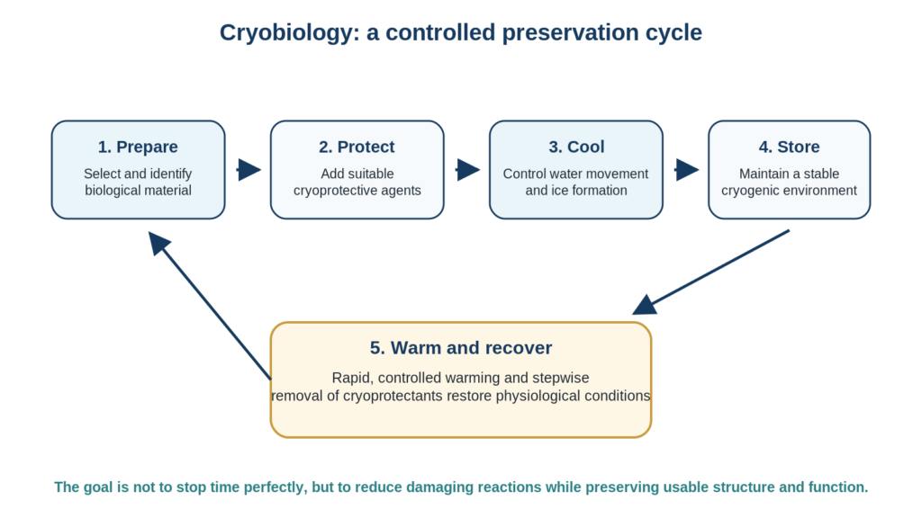
A simplified sequence showing preparation, cryoprotection, cooling, cryogenic storage, warming and recovery. Original educational illustration created for Inside Embryo; schematic and not to scale.
Key Takeaways
- Cryobiology studies how living systems respond to low temperatures; cryopreservation applies that knowledge to preserve biological material.
- Unprotected freezing is usually damaging because ice, concentrated solutes and rapid volume changes can disrupt cells.
- Cryoprotective agents reduce cryoinjury, but they must be used within validated exposure and temperature conditions.
- Slow freezing manages extracellular ice and cell dehydration, whereas vitrification aims to create an ice-free glass-like state.
- Warming is an active part of the method, not merely the reversal of cooling.
- In modern ART, vitrification is widely used for oocytes and embryos, while sperm and tissues may require different approaches.
- Post-warming survival does not by itself prove normal genetic, metabolic, developmental or reproductive competence.
- Safe cryostorage depends on identification, documentation, alarm systems, trained staff, emergency plans and local regulation.
What Cryobiology and Cryopreservation Mean
Cryobiology combines biology, physical chemistry and engineering. It examines how temperature affects water, solutes, membranes, proteins, organelles, tissues and whole biological systems. Cryopreservation is one application of this field: biological material is cooled and stored under conditions designed to maintain later usability.[2,3]
The word preservation should not be interpreted as a guarantee of perfect unchanged function. A specimen may remain viable yet show reduced motility, altered morphology, membrane injury or impaired developmental potential. For this reason, laboratories evaluate outcomes using several measures, such as recovery, survival, motility, fertilisation, embryo development or clinical outcomes, depending on the material and purpose.
Table 1. Essential beginner terminology
| Term | Clear meaning | Why it matters |
| Cryobiology | Study of how low temperatures affect living systems. | Provides the scientific basis for preservation methods. |
| Cryopreservation | Storage of cells or tissues at very low temperature using a controlled method. | Used for reproductive material, research samples and other medical applications. |
| Cryoinjury | Damage caused during cooling, storage or warming. | May arise from ice, osmotic stress, toxicity or thermal effects. |
| Cryoprotective agent (CPA) | A substance that reduces freezing-related injury. | May act inside the cell, outside the cell or both. |
| Vitrification | Conversion of a solution into a glass-like state without significant ice crystallisation. | Widely used for human oocytes and embryos. |
| Warming | Controlled return from cryogenic temperature toward physiological conditions. | Must limit recrystallisation and osmotic injury. |
Table note: CPA = cryoprotective agent.
These terms describe related but different ideas. Cryobiology is the field; cryopreservation is the process; cryoinjury is the damage that the process is designed to prevent or minimise.
Why Ordinary Freezing Can Damage Cells
Cells contain a large amount of water. When an aqueous solution cools below its freezing point, ice tends to form first in the extracellular environment. Because pure ice excludes most dissolved substances, the remaining liquid becomes more concentrated. Water then moves out of the cell in response to this osmotic gradient, causing the cell to shrink.[1,2]
If cooling is too rapid, water may not leave quickly enough and intracellular ice can form. Intracellular crystals may disrupt membranes, organelles and the cytoskeleton. If cooling is too slow, the cell may remain for too long in a highly concentrated extracellular solution and experience excessive dehydration, solute effects and mechanical stress. The classical cryobiological challenge is therefore to avoid both extremes.[1]
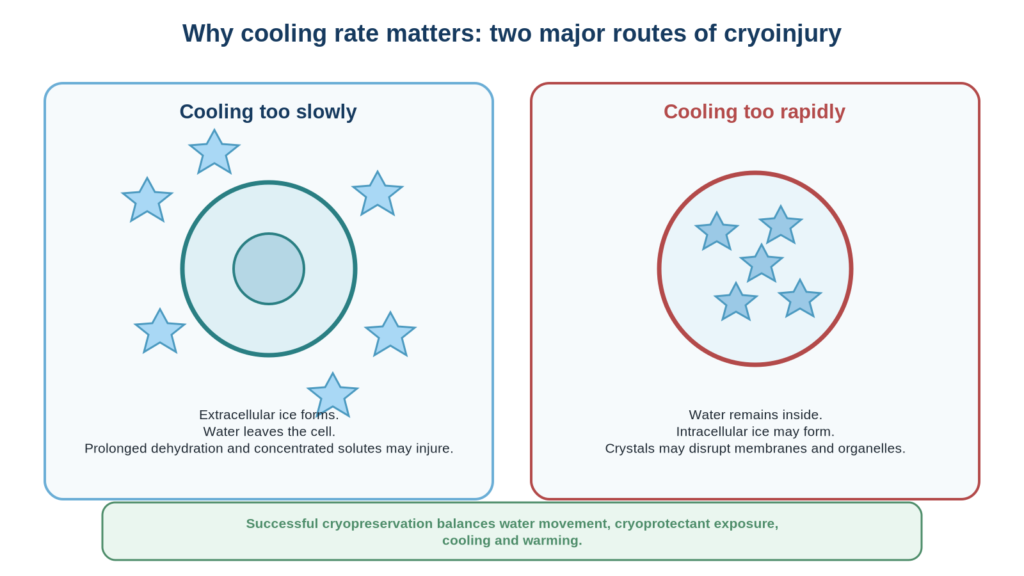
Cooling too slowly may produce prolonged dehydration and concentrated-solute injury; cooling too rapidly may promote intracellular ice. Successful methods balance these competing risks. Original educational illustration created for Inside Embryo; schematic and not to scale.
Osmotic stress and cell-volume changes
Osmosis is the movement of water across a selectively permeable membrane in response to differences in solute concentration. During cryoprotectant addition, cooling, thawing or cryoprotectant removal, the cell may shrink and re-expand. Some change is expected, but abrupt or excessive volume change can injure the plasma membrane and internal structures. Different cell types have different membrane permeability, size, water content and tolerance; a method suitable for spermatozoa cannot simply be transferred to an oocyte or tissue.
Temperature-sensitive cellular structures
Cooling can alter lipid membranes, protein organisation and cytoskeletal structures. The mature oocyte is especially demanding because it is large, contains substantial water and has a temperature-sensitive meiotic spindle. Embryos at different stages also differ in cell number, membrane permeability, blastocoel volume and tolerance. These biological differences help explain why cryopreservation is material-specific rather than a single universal technique.[4,5]
Cryoprotective Agents: Protection with Important Limits
Cryoprotective agents, commonly shortened to CPAs, are compounds added to reduce cryoinjury. They do not make cells immune to freezing. Instead, they alter water movement, ice formation, solution viscosity and the physical behaviour of the preservation medium. Their benefit depends on concentration, temperature, exposure time, cellular permeability and the method used.[2,4]
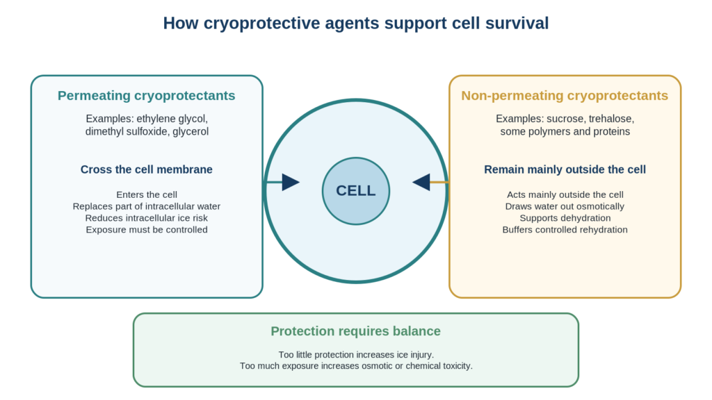
Permeating agents enter cells and replace part of the intracellular water, whereas non-permeating agents act mainly outside the cell to support dehydration and controlled rehydration. Original educational illustration created for Inside Embryo; schematic and not to scale.
Table 2. Main cryoprotectant groups
| Group | Examples | Main role | Important limitation |
| Permeating | Ethylene glycol, dimethyl sulfoxide, glycerol, propylene glycol | Enter cells to replace part of the intracellular water and reduce intracellular ice. | Chemical toxicity and osmotic stress if exposure is excessive or poorly controlled. |
| Non-permeating | Sucrose, trehalose and some larger molecules | Remain mainly outside cells, promote dehydration and assist controlled rehydration. | Excessive extracellular osmolality can over-shrink cells. |
| Macromolecular/supportive components | Selected proteins or polymers in validated media | May support viscosity, membrane protection or solution stability. | Effects vary by formulation and biological material. |
Table note: Examples are educational, not a formulation or protocol.
The key principle is balance. Inadequate CPA exposure may leave too much free water available for ice formation, while excessive exposure can cause osmotic or chemical injury. Laboratories therefore use stepwise handling, defined temperatures and carefully timed transfers within validated methods.
Slow Freezing and Vitrification
Controlled slow freezing and vitrification are the two major conceptual approaches. Both aim to preserve cells, but they manage ice and water differently. Slow freezing permits controlled extracellular ice formation while encouraging water to leave the cell. Vitrification uses concentrated cryoprotective solutions, a very small sample volume and rapid cooling so that the solution becomes a highly viscous glass-like state with minimal crystal formation.[2,4-6]
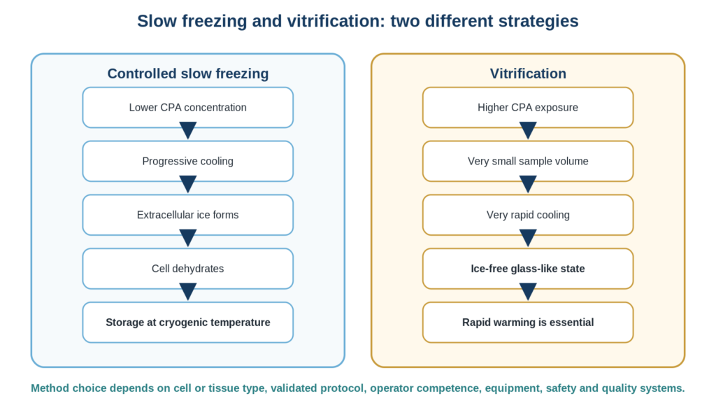
The two strategies differ in cryoprotectant exposure, cooling behaviour and management of ice. Exact procedures must be validated for the biological material and laboratory. Original educational illustration created for Inside Embryo; simplified educational flowchart.
Table 3. Conceptual comparison of slow freezing and vitrification
| Feature | Controlled slow freezing | Vitrification |
| Basic strategy | Progressive cooling with controlled extracellular ice and dehydration. | Very rapid cooling into an ice-free glass-like state. |
| CPA exposure | Usually lower than vitrification, with longer cooling. | Usually higher, but exposure is brief and tightly timed. |
| Ice | Some extracellular ice is expected and managed. | Significant ice formation is avoided. |
| Equipment concept | Often uses programmable or controlled-rate cooling. | Uses specialised carriers and minimum volumes; warming speed is critical. |
| Current reproductive use | Still relevant for some sperm and tissue methods and in established validated systems. | Widely used for human oocytes and embryos in modern ART. |
| Main risks | Intracellular ice, prolonged solute exposure and dehydration injury. | CPA toxicity, osmotic injury, devitrification or recrystallisation during inadequate warming. |
Table note: Specific protocols vary by material, device and laboratory.
A systematic review found better cryosurvival with vitrification than slow freezing for oocytes, cleavage-stage embryos and blastocysts, although the quality of evidence for some clinical outcomes was limited.[5] ASRM guidance considers rapid-cooling vitrification a standard approach for human oocytes and embryos when performed with validated protocols, operator training and quality control.[6] This does not mean vitrification is automatically superior for every cell, tissue or research purpose.
Warming and Recovery
Warming is not a passive afterthought. During warming, a vitrified solution can form ice if it spends too long in a temperature range that permits nucleation or crystal growth. Rapid and controlled warming helps reduce this risk. At the same time, cryoprotectants must be removed without causing sudden water entry and excessive swelling.[2,6]
After warming, the specimen is assessed using criteria appropriate to the material. Sperm assessment may include motility and concentration; oocytes may be evaluated for membrane integrity and morphology; embryos may be observed for cell survival, re-expansion or continued development. A visually intact specimen is encouraging but cannot prove normal molecular function, implantation or live birth.
Applications in Reproductive Science and ART
Cryopreservation has become integral to assisted reproductive technology (ART). It allows reproductive material to be separated in time from collection, fertilisation, embryo transfer or later use. Common applications include sperm banking before treatment or surgery, fertility preservation, donor programmes, storage of supernumerary embryos, planned oocyte cryopreservation, and preservation of ovarian or testicular tissue in selected settings.[7-10]
For a broader overview of gametes, fertilisation and reproductive function, see Human Reproductive Biology.
Spermatozoa
Sperm cryopreservation is widely used because sperm cells are small and can often be stored successfully using established methods. Nevertheless, freezing and warming may reduce motility and membrane function in a proportion of cells. The World Health Organization laboratory manual includes standardised approaches relevant to semen processing and cryopreservation, while interpretation and clinical use must remain context-specific.[8]
Oocytes and embryos
Modern vitrification has transformed oocyte and embryo banking. It supports fertility preservation, donor-oocyte programmes, frozen embryo transfer, embryo accumulation in selected clinical strategies and safe postponement of transfer when clinically appropriate. ESHRE and ASRM sources recognise oocyte and embryo cryopreservation as established components of reproductive medicine, but outcomes remain influenced by age, embryo or oocyte quality, laboratory performance and clinical factors.[5-7,9]
Developmental context is available in Human Embryology: 8 Essential Stages from Fertilization to Organ Formation.
Reproductive tissues
Ovarian and testicular tissues are more complex than isolated cells because they contain multiple cell types, extracellular matrix, blood vessels and spatial relationships. Ovarian tissue cryopreservation is an established fertility-preservation option in selected circumstances, while some testicular tissue approaches—particularly for prepubertal patients—remain specialised or experimental depending on intended use and jurisdiction.[9]
Quality, Safety and Cryostorage
Once a specimen has been cryopreserved, safe storage becomes a long-term quality responsibility. Cryostorage systems commonly use liquid nitrogen, which is approximately -196 °C in the liquid phase. At sufficiently low temperature, molecular movement and damaging reactions are greatly reduced, but this does not remove the need for active monitoring, inventory control and emergency planning.[7,11]
Current ESHRE good-practice recommendations emphasise restricted access, ventilation, low-oxygen alarms, continuous monitoring of storage tanks, backup capacity, personal protective equipment, staff training, validated transport containers, documentation, permanent labelling, inventory checks and identity verification.[7] ASRM also recommends rigorous policies for tank management and emergency preparedness because reproductive specimens may be irreplaceable.[11,12]
Liquid nitrogen can displace oxygen and cause cold burns, so cryostorage rooms require risk assessment and trained personnel. Storage requirements, maximum storage periods, consent rules, infectious-risk management and disposition procedures vary by country. A global article can describe principles, but each laboratory must follow current local law and accreditation requirements.
Common Misunderstandings, Limitations and Evidence Status
Table 4. Common misunderstandings and accurate interpretation
| Misunderstanding | More accurate interpretation |
| “Freezing stops all damage forever.” | Very low temperature greatly slows reactions, but risk remains during handling, transport, warming, tank failure or temperature excursion. |
| “A cell that survives warming is completely normal.” | Survival confirms basic integrity, not full genetic, metabolic, developmental or reproductive competence. |
| “Vitrification means no risk.” | Vitrification reduces ice formation but introduces osmotic, toxic, handling and warming-related risks. |
| “One protocol works for every specimen.” | Cell size, permeability, water content, developmental stage and tissue structure require different validated methods. |
| “Storage duration alone determines outcome.” | Material quality at freezing, method, temperature stability, warming, age and clinical factors also matter; long-term evidence differs by specimen type. |
Evidence is strongest for routine sperm, oocyte and embryo cryopreservation in established ART programmes. Evidence is less uniform for some tissue approaches, novel devices, automated systems, repeated cryopreservation, very long storage intervals and emerging preservation technologies. Professional recommendations may combine published evidence with expert consensus when trials are limited.[5-7]
Practical Relevance for Learners and Laboratories
For a beginner, the most useful mental model is a chain of controlled stresses. The specimen is first identified and assessed, then exposed to protective media, cooled by a validated method, stored below a critical temperature, warmed quickly enough to prevent damaging ice formation, and returned gradually to physiological conditions. Every link in this chain affects the final outcome.
For an IVF laboratory, technical skill must be supported by a quality management system. Training, competency assessment, witnessing or double-check systems, media and device validation, environmental control, traceability, equipment qualification, maintenance, alarms, incident reporting and emergency drills are not administrative extras. They are part of the scientific reliability of cryopreservation.[7,11,12]
Learners should understand the principles before studying a specific commercial kit or laboratory SOP. The theory explains why timing matters, why cells shrink and recover, why minimum volume is important in vitrification, why warming must be rapid, and why deviations can affect outcomes.
Continue with the IVF Laboratory Science Learning Hub for related laboratory foundations.
Frequently Asked Questions
Is cryobiology the same as cryopreservation?
No. Cryobiology is the broader science of low-temperature effects on living systems. Cryopreservation is a practical method developed from that science.
Does liquid nitrogen freeze cells directly?
Sometimes specimens are placed into or above liquid nitrogen after preparation, but survival depends on the full validated method, including cryoprotectants, sample volume, cooling and warming.
Why are cryoprotectants needed?
They reduce damaging ice formation and help control water movement. They can also be harmful if concentration, temperature or exposure are not controlled.
What is the difference between thawing and warming?
The terms are often used informally, but vitrified specimens are usually described as being warmed because they are intended to remain ice-free rather than undergo conventional melting of ice.
Are vitrified embryos guaranteed to survive?
No. Modern vitrification can achieve high survival in competent laboratories, but survival is not guaranteed and does not guarantee implantation, pregnancy or live birth.
Can frozen reproductive material be stored indefinitely?
Biological reactions are extremely limited at stable cryogenic temperatures, but legal storage limits, consent, tank management, funding and long-term evidence vary by jurisdiction and specimen.
Is cryopreservation safe for laboratory staff?
It can be performed safely with training, ventilation, oxygen monitoring, protective equipment, restricted access and emergency procedures. Liquid nitrogen presents cold-burn and oxygen-displacement hazards.
Conclusion
This introduction to cryobiology shows that successful preservation depends on controlling physical and biological change rather than simply reaching a low temperature. Ice formation, water movement, solute concentration, cryoprotectant exposure, cooling rate and warming rate interact with the unique properties of each cell or tissue.
In reproductive medicine, cryobiology makes sperm, oocyte, embryo and tissue banking possible and supports many modern ART pathways. However, post-warming appearance or survival is only one part of outcome assessment. Reliable practice requires validated methods, trained staff, traceability, monitoring, safety systems, current guidance and transparent acknowledgement of uncertainty.
Explore the Cryobiology Learning Hub for future supporting lessons and resources.
More Articles on Inside Embryo
- Introduction to Cellular Biology: Structure, Function and the Life of a Cell
- Human Reproductive Biology: 7 Concepts from Gametes to Implantation
- Human Embryology: 8 Essential Stages from Fertilization to Organ Formation
- Cryobiology Learning Hub
- IVF Laboratory Science Learning Hub
References
1. Mazur P. Freezing of living cells: mechanisms and implications. Am J Physiol. 1984;247(3 Pt 1):C125-C142. doi:10.1152/ajpcell.1984.247.3.C125. DOI or official source
2. Pegg DE. Principles of cryopreservation. Methods Mol Biol. 2007;368:39-57. doi:10.1007/978-1-59745-362-2_3. DOI or official source
3. Jang TH, Park SC, Yang JH, et al. Cryopreservation and its clinical applications. Integr Med Res. 2017;6(1):12-18. doi:10.1016/j.imr.2016.12.001. DOI or official source
4. Konc J, Kanyó K, Kriston R, Somoski B, Cseh S. Cryopreservation of embryos and oocytes in human assisted reproduction. Biomed Res Int. 2014;2014:307268. doi:10.1155/2014/307268. DOI or official source
5. Rienzi L, Gracia C, Maggiulli R, et al. Oocyte, embryo and blastocyst cryopreservation in ART: systematic review and meta-analysis comparing slow-freezing versus vitrification. Hum Reprod Update. 2017;23(2):139-155. doi:10.1093/humupd/dmw038. DOI or official source
6. Practice Committees of ASRM and SRBT. A review of best practices of rapid-cooling vitrification for oocytes and embryos: a committee opinion. Fertil Steril. 2021. DOI or official source
7. ESHRE Good Practice in the IVF Lab Working Group. ESHRE recommendations on Good Practice in the IVF laboratory. Hum Reprod. 2026;deag096. doi:10.1093/humrep/deag096. DOI or official source
8. World Health Organization. WHO laboratory manual for the examination and processing of human semen. 6th ed. Geneva: WHO; 2021. DOI or official source
9. ESHRE Guideline Group on Female Fertility Preservation, Anderson RA, Amant F, et al. ESHRE guideline: female fertility preservation. Hum Reprod Open. 2020;2020(4):hoaa052. doi:10.1093/hropen/hoaa052. DOI or official source
10. Estudillo E, Jiménez A, Bustamante-Nieves PE, Palacios-Reyes C, Velasco I, López-Ornelas A. Cryopreservation of gametes and embryos and their molecular changes. Int J Mol Sci. 2021;22(19):10864. doi:10.3390/ijms221910864. DOI or official source
11. Practice Committees of ASRM, SART and SRBT. Cryostorage of reproductive tissues in the in vitro fertilization laboratory: a committee opinion. Fertil Steril. 2020;114(3):486-491. DOI or official source
12. Practice Committee of ASRM. Development of an emergency plan for in vitro fertilization programs: a committee opinion. Fertil Steril. 2021. DOI or official source
About the Author
Manoj Kumar K is an Embryologist and the founder of Inside Embryo, an independent educational platform focused on assisted reproduction, human embryology and IVF laboratory science. His work combines laboratory perspective with evidence-based scientific education for students, laboratory professionals, clinicians, researchers and interested readers worldwide. Read the full author profile at:
Editorial Transparency and Corrections
Scientific information may change as evidence, technology and professional guidance develop. Readers may report a suspected factual or reference error through https://insideembryo.com/contact/. Publication and update dates should be revised when material changes are made. AI and digital tools are not scientific authorities; the responsible author must verify and approve the final article before publication.
Educational Disclaimer
Educational Disclaimer: This article is intended for scientific and educational purposes only. It does not replace professional medical advice, clinical judgement, institutional policies, validated laboratory protocols, manufacturer instructions, regulatory requirements or formal professional training. Laboratory and clinical procedures should be performed only by appropriately qualified personnel. Laws, standards and professional requirements may differ between countries and may change over time. Readers should consult current official guidance and qualified professionals for clinical, laboratory or personal health decisions.

Nice