From ovulation and sperm transport to pronuclei and the first cell division.
A clear step-by-step explanation of how a sperm reaches an oocyte, how the two gametes fuse, and how a one-cell zygote begins early development.
| CATEGORY – IVF & Fertility Basics | READING TIME ~12-14 minutes | AUTHOR – Manoj Kumar K, Embryologist |
| PUBLISHED – 5 October 2026 | REVIEW – 5 October 2026 | PUBLISHER – Inside Embryo by Aurion |
Fertilisation is often shortened to the phrase “sperm meets egg.” Biologically, it is much more than a single moment. It is a coordinated sequence in which a sperm must reach the oocyte, become functionally ready to fertilise, pass through the oocyte’s surrounding layers, bind and fuse with the oocyte membrane, and trigger activation of the oocyte. Only after these steps can the maternal and paternal genetic material prepare for the first embryonic cell division. [1,5,7]
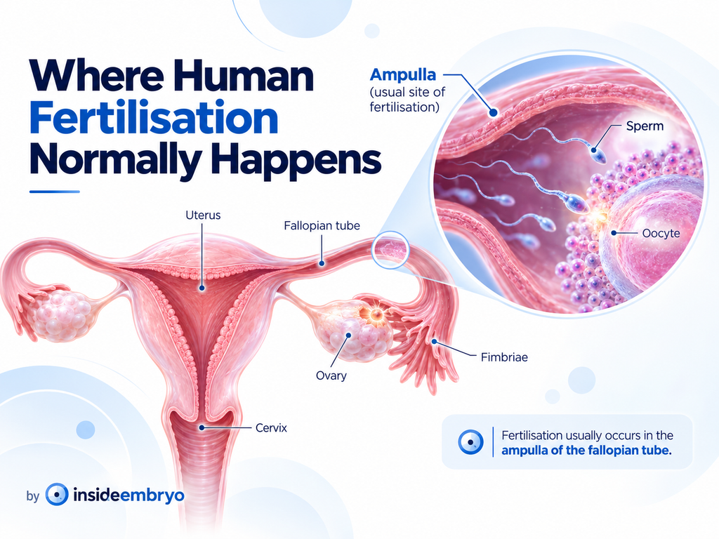
In unassisted conception, fertilisation usually occurs in the fallopian tube – most commonly in the ampullary region – after ovulation. The uterus is normally the site where the developing embryo later implants, not the usual site where sperm and oocyte first fuse. [1,3,6]
| In one sentence Human fertilisation is the multistep process in which a functionally prepared sperm reaches and fuses with a mature oocyte, activates it, contributes paternal genetic material, and helps form the one-cell zygote that can begin cleavage. |
Table of Contents
What is human fertilisation?
Human fertilisation is the process by which the male gamete (sperm) and female gamete (oocyte) unite and initiate development of a new one-cell zygote. A mature human sperm and a mature human oocyte are haploid cells, meaning each normally carries 23 chromosomes. After successful fertilisation, the resulting zygote normally contains 46 chromosomes arranged in 23 pairs. [6]
The process is not simply the physical entry of a sperm. Successful fertilisation requires several biological events to occur in the correct sequence: sperm preparation, interaction with the oocyte coverings, gamete membrane fusion, oocyte activation, prevention of additional sperm entry, completion of the oocyte’s second meiotic division, formation of the maternal and paternal pronuclei, and preparation for the first mitotic division. [5,7]
Where does fertilisation normally happen?
In unassisted conception, sperm travel from the vagina through the cervix and uterus toward the fallopian tubes. After ovulation, the oocyte is captured by the fimbrial end of the tube and moves into the tubal lumen. The ampulla – the wider middle portion of the fallopian tube – is the usual site of fertilisation. [1,3,6]
| Important to know Fertilisation and implantation are different events. Fertilisation usually takes place in the fallopian tube. Implantation occurs later, after the developing embryo reaches the uterus and attaches to the endometrium. |
Fertilisation at a glance
| Stage | What happens | Why it matters |
| 1. Ovulation | A mature oocyte is released from the ovary and enters the fallopian tube. | The oocyte must be present while viable sperm are available. |
| 2. Sperm transport | Sperm move through the cervix, uterus and toward the fallopian tube. | Only sperm that reach the appropriate site can participate in fertilisation. |
| 3. Capacitation | Sperm undergo functional changes within the female reproductive tract. | These changes help sperm develop the ability to fertilise the oocyte. |
| 4. Cumulus interaction | Sperm pass through the cells surrounding the oocyte. | This brings sperm to the zona pellucida around the oocyte. |
| 5. Zona interaction / acrosome reaction | A fertilising sperm undergoes acrosomal changes and penetrates the zona pellucida. | This allows the sperm to reach the oocyte plasma membrane. |
| 6. Membrane fusion | The sperm membrane binds and fuses with the oolemma. | The sperm can deliver its genetic material and activation signal. |
| 7. Oocyte activation | Calcium-dependent signalling begins and cortical granules are released. | The oocyte resumes development and mechanisms limit entry of additional sperm. |
| 8. Pronuclear stage | The oocyte completes meiosis II and maternal and paternal pronuclei form. | The two parental genomes prepare for the first embryonic division. |
| 9. First cleavage | Pronuclear membranes break down and chromosomes organise for mitosis. | The one-cell zygote begins dividing into early embryonic cells. |
See This Topic on Instagram
Explore our related Instagram post for a quick visual overview.
1. Ovulation: the oocyte enters the fallopian tube
Before fertilisation can occur naturally, an oocyte must be released from the ovary. During ovulation, the dominant follicle releases the oocyte-cumulus complex near the fimbrial end of the fallopian tube. The fimbriae and tubal movements help guide the complex into the tube. [3,6]
The ovulated human oocyte is usually a secondary oocyte arrested at metaphase II of meiosis. It has not yet completed its final meiotic division. Completion of meiosis II is normally triggered only after fertilisation activates the oocyte. [6,7]
| Timing matters Patient guidance commonly describes the ovulated oocyte as remaining fertilisable for roughly 24 hours, while sperm may remain viable in the female reproductive tract for about 3 days and sometimes up to 5 days. This is why intercourse before ovulation can still result in fertilisation. [2] |
2. The sperm journey: from ejaculation to the fallopian tube
After ejaculation into the vagina, sperm must move through the cervical mucus, enter the uterine cavity and reach the fallopian tube. This journey is not simply a swimming race. Cervical mucus, uterine and tubal contractions, fluid movement, sperm motility and the timing of ovulation all influence transport. Many sperm begin the journey, but only a small fraction approach the site where an oocyte may be present. [5,7]
Around ovulation, cervical mucus becomes more favourable for sperm passage. Sperm that reach the upper reproductive tract are exposed to conditions that support the next essential process: capacitation. [2,5]
3. Capacitation: preparing sperm to fertilise
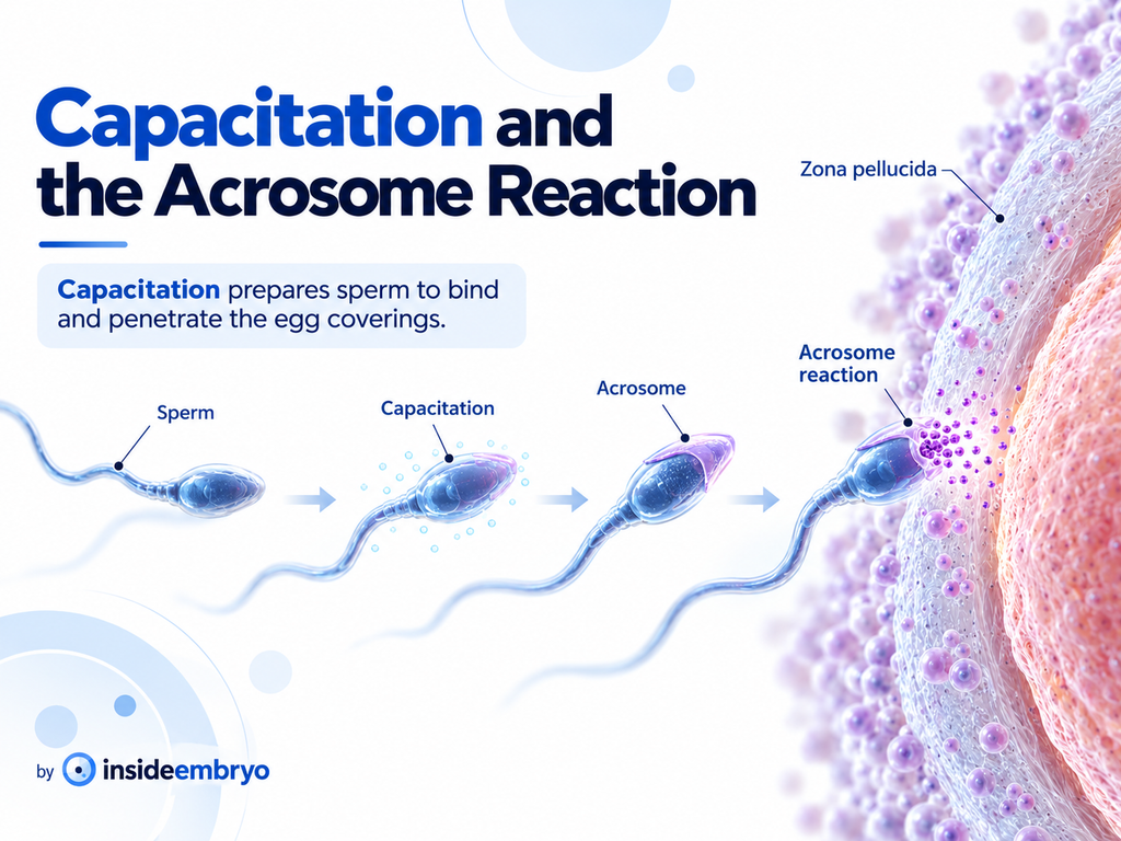
Freshly ejaculated sperm are motile, but they are not yet fully competent to fertilise an oocyte. In the female reproductive tract they undergo a collection of biochemical and membrane changes called capacitation. These changes alter the sperm surface, signalling pathways and movement pattern, allowing the sperm to respond appropriately to the environment around the oocyte. [5,7]
Capacitation is not a visible “switch” that happens at one exact location. It develops over time as sperm interact with the reproductive tract. One consequence is a more forceful, asymmetric pattern of movement called hyperactivation, which helps sperm negotiate the complex environment around the oocyte and its coverings. [5,7]
| A useful way to think about capacitation The sperm may already be moving after ejaculation, but capacitation is part of what makes it functionally ready to complete the later steps of fertilisation. |
4. Reaching the oocyte: cumulus cells and the zona pellucida
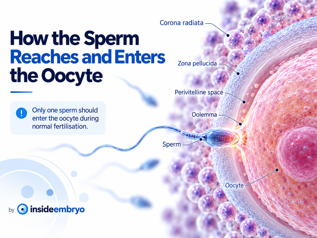
The ovulated oocyte is not exposed directly to the fallopian-tube fluid. It is surrounded by cumulus cells embedded in a specialised extracellular matrix. Beneath these cells lies the zona pellucida, a glycoprotein-rich coat surrounding the oocyte. A sperm capable of fertilisation must navigate the cumulus layer and then interact successfully with the zona pellucida. [5,7]
The zona pellucida is not just a passive shell. It helps organise sperm-oocyte interactions, protects the oocyte and early embryo, and later participates in mechanisms that prevent additional sperm from fertilising an already fertilised oocyte. [5,7]
5. The acrosome reaction and penetration of the zona pellucida
The acrosome is a specialised structure over the front of the sperm head. During the acrosome reaction, the sperm undergoes major membrane changes and releases or exposes molecules needed for the later stages of zona penetration and oocyte interaction. The reaction is essential for a sperm to become capable of fusing with the oocyte membrane. [5,7]
Older diagrams often show the zona pellucida as the single trigger for the acrosome reaction. Current research shows a more complex picture: in humans and other mammals, acrosomal changes may begin before direct zona binding, within the cumulus environment, or during zona interaction. The precise timing is therefore more dynamic than the classic “touch zona, then react” model suggests. [7]
After the sperm has completed the necessary acrosomal changes, it can penetrate the zona pellucida and enter the perivitelline space – the narrow space between the zona and the oocyte membrane.
6. Sperm-oocyte membrane fusion: the decisive contact
Once an appropriately prepared sperm reaches the oocyte plasma membrane, called the oolemma, specific proteins on the sperm and oocyte support recognition, adhesion and membrane fusion. One well-established interaction involves the sperm protein IZUMO1 and its oocyte receptor JUNO, although fertilisation depends on a wider network of molecules rather than a single protein pair. [7,8]
Membrane fusion allows the sperm to deliver its nucleus and sperm-derived factors into the oocyte cytoplasm. This is the event that initiates oocyte activation and starts the transition from an unfertilised oocyte toward a zygote.
| The “fastest sperm wins” idea is too simple Sperm selection in vivo is influenced by transport through the reproductive tract, capacitation, motility, cumulus interaction, zona penetration and molecular compatibility at the oocyte membrane. Fertilisation is therefore a multistep biological selection process, not just a straight swimming contest. |
7. Oocyte activation: switching the egg into developmental mode
Sperm-oocyte fusion triggers a series of calcium-dependent signals inside the oocyte. These calcium oscillations are central to oocyte activation. Activation allows the oocyte to resume and complete meiosis II, reorganise its cellular machinery, and begin the biochemical programme needed for early embryo development. [4,5,7]
If normal activation does not occur, the oocyte may fail to complete fertilisation even when a sperm has reached or entered it. This is one reason fertilisation cannot be reduced to sperm entry alone. [4]
8. Blocking polyspermy: why only one sperm should fertilise the oocyte
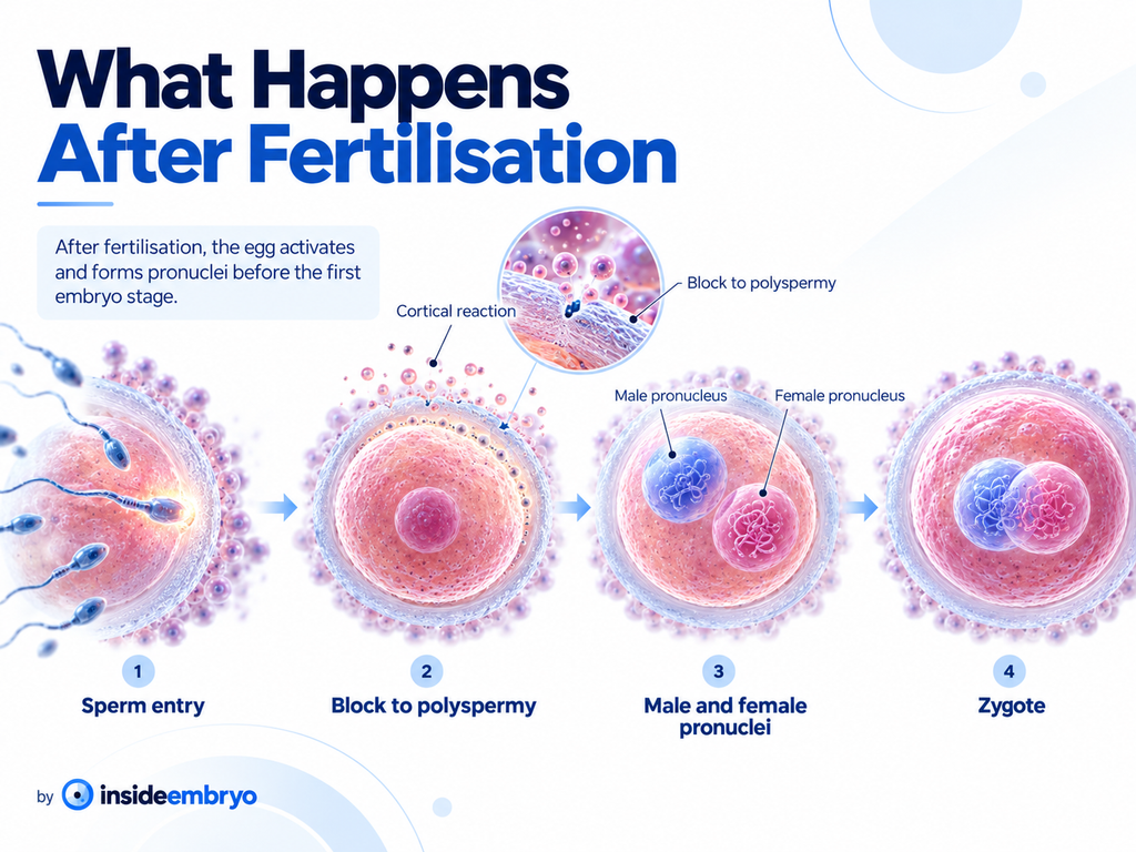
Normal human fertilisation requires contribution from one sperm. Entry or fusion of additional sperm – polyspermy – creates an abnormal chromosome complement and is generally incompatible with normal development. The oocyte therefore activates mechanisms that rapidly make additional fertilisation less likely. [5,7]
A major part of this response is the cortical reaction. Cortical granules located beneath the oocyte membrane release their contents after activation. These products alter the zona pellucida and the oocyte surface, helping prevent other sperm from binding, penetrating or fusing. Modern research shows that the block to polyspermy involves coordinated changes at both the zona pellucida and oolemma. [7,9]
| Why this matters A normal fertilisation event is not “more sperm is better.” The biological goal is one sperm genome plus one oocyte genome, followed by accurate chromosome organisation for the first cell division. |
9. Completion of meiosis II and formation of the pronuclei
After activation, the secondary oocyte completes meiosis II and extrudes the second polar body. The maternal chromosomes are organised into the female pronucleus. At the same time, the highly condensed sperm chromatin decondenses and becomes organised into the male pronucleus. [5,6]
For a period, the maternal and paternal genetic material remain in two separate pronuclei within the same cell. In an IVF laboratory, seeing two pronuclei – often written as 2PN – at the expected assessment time is the classic morphological sign of normal fertilisation. However, a 2PN appearance does not guarantee that an embryo will continue to develop normally. [3,6]
As the cell prepares for its first mitotic division, the pronuclear envelopes break down and the parental chromosomes become organised on the first mitotic spindle. The genetic material is therefore not simply “mixed together” the moment the sperm enters the oocyte; it undergoes a carefully regulated sequence before the first cleavage. [5,6]
What changes at fertilisation?
| Before fertilisation | After successful fertilisation |
| The oocyte is a secondary oocyte arrested at metaphase II. | Oocyte activation allows completion of meiosis II. |
| The oocyte normally carries 23 maternal chromosomes. | Maternal chromosomes form the female pronucleus. |
| The sperm normally carries 23 paternal chromosomes. | Sperm chromatin decondenses and forms the male pronucleus. |
| The oocyte must still block entry of extra sperm. | Cortical and membrane changes reduce the risk of polyspermy. |
| No embryonic cleavage has begun. | The one-cell zygote prepares for its first mitotic division. |
What happens after fertilisation?
Fertilisation creates the one-cell zygote, but pregnancy requires several further stages. The zygote begins cleavage – repeated cell divisions without a major increase in overall size – while moving through the fallopian tube toward the uterus. It progresses through early cleavage stages, compaction and morula formation, and then forms a blastocyst. The blastocyst later hatches from the zona pellucida and can begin implantation in the uterine lining. [1,3,6]
| Approximate stage | What is happening |
| Day 0 | Fertilisation and formation of the one-cell zygote. |
| Around Day 1 | First cleavage produces two early embryonic cells. |
| Around Day 2-3 | Further cleavage creates several blastomeres. |
| Around Day 4 | Compaction leads toward the morula stage. |
| Around Day 5-6 | A blastocyst forms as the embryo reaches or is within the uterus. |
| Around Day 6 onward | After hatching, attachment and implantation can begin. |
| These timings are approximate Human development varies, and exact timing cannot be observed directly in natural conception. IVF laboratories can observe embryos at defined time points, but even there, normal embryos do not all develop at exactly the same speed. |
Fertilisation is not the same as implantation or pregnancy
Fertilisation is the union and activation process involving sperm and oocyte. Implantation happens later, when a developing blastocyst attaches to and begins interacting with the uterine lining. A fertilised oocyte must therefore continue developing, reach the uterus and implant successfully before an ongoing pregnancy can be established. [1]
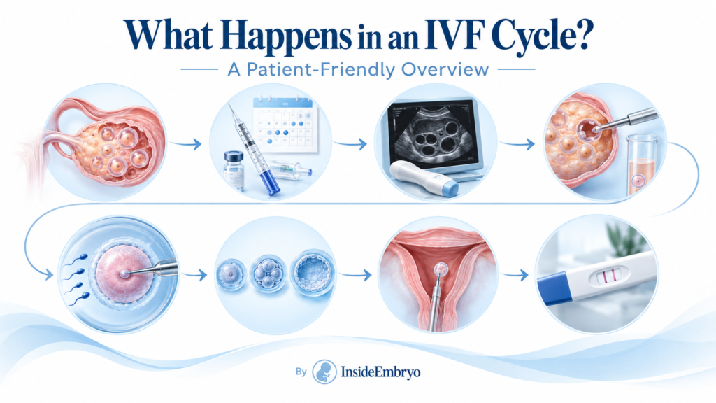
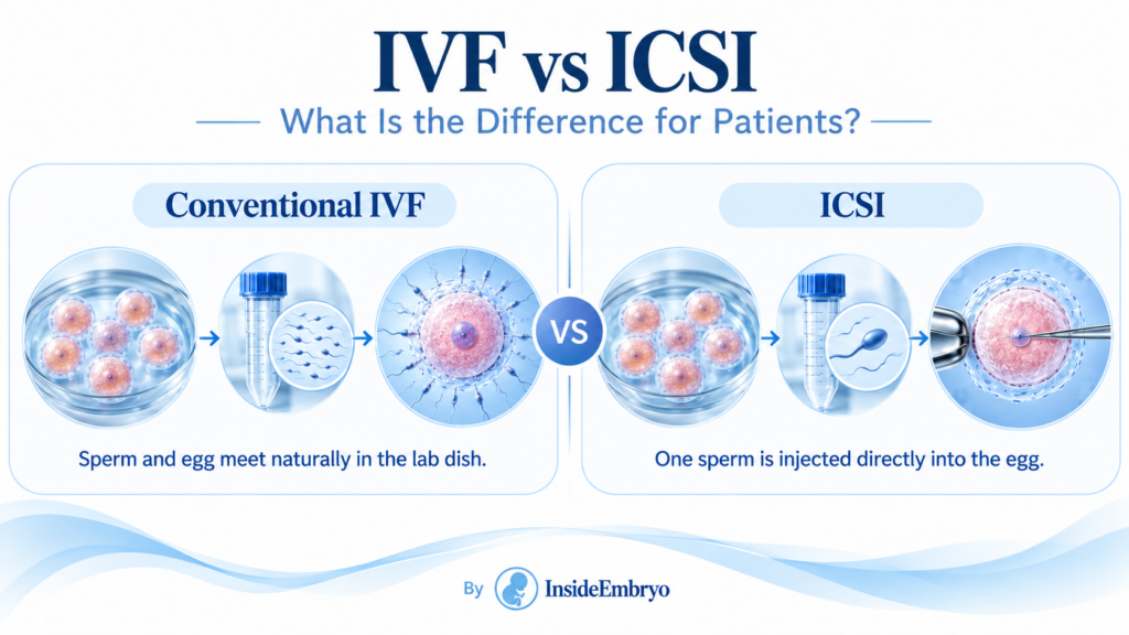
Natural fertilisation vs conventional IVF vs ICSI
The core biology of oocyte activation, pronuclear formation and early cell division is relevant to all three pathways, but the way sperm and oocyte are brought together differs.
| Feature | Unassisted fertilisation | Conventional IVF | ICSI |
| Where sperm and oocyte meet | Usually in the fallopian tube. | In a laboratory culture dish. | In the laboratory at a micromanipulation workstation. |
| How sperm reaches the oocyte | Sperm travels through the reproductive tract and penetrates the oocyte coverings. | Prepared sperm are placed around the oocyte-cumulus complex and sperm-oocyte interaction occurs in culture. | An embryologist selects one sperm and injects it directly into a mature oocyte. |
| Steps bypassed | None of the natural sperm-entry steps are bypassed. | Transport through the female reproductive tract is bypassed, but sperm still interacts with the oocyte coverings. | Transport, cumulus/zona penetration and membrane-entry steps are largely bypassed by direct injection. |
| What still must happen | Oocyte activation, pronuclear formation and early development. | Oocyte activation, pronuclear formation and early development. | The injected oocyte still has to activate, form pronuclei and support early development. |
| Connection to IVF ICSI can bypass important sperm-entry barriers, but it does not guarantee normal fertilisation. Even after injection, the oocyte still has to activate and complete the biological events of fertilisation. |
Why might fertilisation not occur?
Fertilisation can fail for many different reasons, and one unsuccessful event does not automatically identify a single cause. Possible factors include:
- Timing: viable sperm and a fertilisable oocyte may not be present in the same place at the same time.
- Tubal factors: blockage or severe tubal damage can prevent sperm and oocyte from meeting naturally.
- Sperm factors: very low sperm number, reduced motility, abnormal function, impaired capacitation or problems with acrosomal or membrane-fusion steps may reduce fertilising ability.
- Oocyte factors: an oocyte may be immature, biologically compromised, or unable to activate normally after sperm interaction.
- Sperm-oocyte interaction defects: fertilisation can be disrupted at the cumulus, zona pellucida, oolemma or activation stages.
- Genetic or molecular factors: rare variants affecting proteins needed for gamete recognition, fusion or activation can contribute to fertilisation failure.
- Unexplained factors: sometimes the exact reason cannot be identified from routine clinical testing.
In fertility treatment, the interpretation depends on the full clinical history, semen findings, number and maturity of oocytes, previous fertilisation results, and the laboratory method used. A fertility specialist and embryology team can explain what a particular result may mean for an individual cycle.
Myth vs fact
| Myth | Fact |
| Myth: The fastest sperm is automatically the one that fertilises the oocyte. | Fact: Speed is only one small part of a complex process involving transport, capacitation, motility pattern, cumulus and zona interaction, and molecular recognition at the oocyte membrane. |
| Myth: Human fertilisation normally happens in the uterus. | Fact: It usually occurs in the fallopian tube, most commonly in the ampullary region. The uterus becomes important later for implantation. |
| Myth: Once one sperm enters, pregnancy is guaranteed. | Fact: Fertilisation is only the beginning. The embryo must continue developing, reach the uterus, implant and progress through many later stages. |
| Myth: More sperm around the oocyte always improves fertilization | Fact: Normal fertilisation requires one sperm to contribute its genome. The oocyte actively changes after fertilisation to reduce the chance of polyspermy. |
| Myth: Fertilisation and implantation are the same event. | Fact: Fertilisation forms the zygote; implantation occurs several days later when the blastocyst attaches to the endometrium. |
Frequently asked questions
Can you feel fertilisation happening?
No reliable physical sensation tells you that fertilisation has occurred. Symptoms immediately after intercourse or ovulation cannot confirm whether a sperm and oocyte have fused.
How long can an oocyte be fertilised after ovulation?
Patient guidance commonly describes the oocyte as remaining fertilisable for about 24 hours after ovulation, although biology varies. [2]
How long can sperm survive in the female reproductive tract?
Sperm can commonly survive for about 3 days and sometimes up to 5 days when conditions are favourable. [2]
Can two sperm fertilise one oocyte?
Polyspermy can occur abnormally, but normal fertilisation activates mechanisms designed to prevent additional sperm entry. Polyspermic fertilisation usually creates an abnormal chromosome complement and does not support normal development.
What does 2PN mean in an IVF laboratory?
2PN means two pronuclei are visible after fertilisation assessment – one maternal and one paternal pronucleus. It is the classic morphological sign of normal fertilisation, but it does not guarantee later embryo development.
Does fertilisation mean an embryo is genetically normal?
No. Fertilisation shows that key early events occurred, but chromosome abnormalities and other developmental problems can still be present.
Can fertilisation happen naturally without fallopian tubes?
Unassisted fertilisation normally requires sperm and oocyte to meet in the reproductive tract, usually the fallopian tube. IVF bypasses the tubes by retrieving oocytes and attempting fertilisation in the laboratory.
Is ICSI the same as natural fertilisation?
No. ICSI bypasses several sperm-entry steps by placing a sperm directly into the oocyte cytoplasm. The oocyte must still activate, form pronuclei and support early embryo development.
Key takeaways
- Human fertilisation is a sequence of coordinated events, not a single instant.
- In unassisted conception, fertilisation usually occurs in the ampulla of the fallopian tube.
- Sperm must undergo capacitation before it becomes fully competent to fertilise an oocyte.
- The fertilising sperm must navigate the cumulus cells and zona pellucida before reaching the oolemma.
- Sperm-oocyte fusion activates the oocyte and triggers mechanisms that reduce the chance of polyspermy.
- The oocyte completes meiosis II and the maternal and paternal pronuclei form before the first cleavage.
- A normally fertilised zygote usually contains 46 chromosomes – 23 from the oocyte and 23 from the sperm.
- Fertilisation does not equal implantation, pregnancy or live birth; many later biological steps still have to occur.
- Conventional IVF and ICSI change how sperm and oocyte are brought together, but normal activation and early development are still required.
Beginner glossary
| Term | Patient-friendly meaning |
| Oocyte | The female reproductive cell; commonly called the egg. |
| Spermatozoon / sperm | The male reproductive cell that carries paternal genetic material. |
| Gamete | A reproductive cell – sperm or oocyte – that normally carries 23 chromosomes. |
| Ovulation | Release of an oocyte from an ovarian follicle. |
| Fallopian tube | The tube connecting the region near the ovary with the uterus; the usual site of natural fertilisation. |
| Ampulla | The wider part of the fallopian tube where fertilisation most commonly occurs. |
| Cumulus cells | Cells surrounding the ovulated oocyte within a specialised extracellular matrix. |
| Zona pellucida | The glycoprotein-rich coat surrounding the oocyte and early embryo. |
| Capacitation | Functional changes that sperm undergo in the female reproductive tract before they become fully competent to fertilise. |
| Acrosome | A specialised structure over the front of the sperm head involved in fertilisation. |
| Acrosome reaction | Membrane and molecular changes in the sperm that help it penetrate the oocyte coverings and become fusion competent. |
| Oolemma | The plasma membrane of the oocyte. |
| Oocyte activation | Calcium-dependent cellular changes triggered by fertilisation that allow the oocyte to complete meiosis and begin development. |
| Polyspermy | Abnormal fertilisation involving more than one sperm. |
| Pronucleus | The temporary maternal or paternal nucleus seen after fertilisation before the first mitotic division. |
| Zygote | The one-cell stage formed after fertilisation. |
| Cleavage | Early mitotic cell divisions of the zygote/embryo without a major increase in overall size. |
| Blastocyst | A later preimplantation embryo stage containing a fluid-filled cavity and distinct cell populations. |
| Implantation | The process by which a blastocyst attaches to and begins interacting with the uterine lining. |
| ICSI | Intracytoplasmic sperm injection: injection of one sperm directly into a mature oocyte during an IVF cycle. |
Suggested related reading on Inside Embryo
- What Happens in an IVF Cycle? A Patient-Friendly Overview
- IVF vs ICSI: What Is the Difference for Patients?
- What Is an Oocyte? A Beginner-Friendly Explanation
- What Happens During Fertilisation in an IVF Laboratory?
- Embryo Development: From Fertilisation to Blastocyst
- What Does an Embryologist Do During Your IVF Cycle?
- Fertility Glossary – sperm, oocyte, fertilisation, zygote and embryo terms
About the author
Manoj Kumar K, Embryologist
Founder and author of Inside Embryo, an educational platform focused on fertility, assisted reproduction, human embryology and IVF laboratory science. The aim is to make complex reproductive science clear, structured and responsible for patients, learners and fertility professionals.
Medical disclaimer
| Educational information only This article provides general education about normal human fertilisation. It does not diagnose infertility, predict an individual chance of conception, recommend a fertility treatment, or replace personalised advice from a fertility specialist or other qualified healthcare professional. Fertilisation biology and fertility problems can vary between individuals, and clinical decisions should be based on a complete medical assessment. For urgent symptoms or concerns, seek appropriate medical care. |
Read the full Inside Embryo Medical Disclaimer
References and trusted resources
1. American College of Obstetricians and Gynecologists (ACOG). How Your Fetus Grows During Pregnancy. Fertilisation and early development. Open source
2. American College of Obstetricians and Gynecologists (ACOG). Fertility Awareness-Based Methods of Family Planning. Ovulation, sperm survival and the fertile window. Open source
3. Human Fertilisation and Embryology Authority (HFEA). In vitro fertilisation (IVF). Patient information on fertilisation outside the body and embryo development. Open source
4. Human Fertilisation and Embryology Authority (HFEA). Treatment add-ons: artificial egg activation. Background on oocyte activation. Open source
5. Georgadaki K, Khoury N, Spandidos DA, Zoumpourlis V. The molecular basis of fertilization. International Journal of Molecular Medicine. 2016;38(4):979-986. doi:10.3892/ijmm.2016.2723. Open source
6. StatPearls. Embryology, Week 1. NCBI Bookshelf. Overview of fertilisation, pronuclei, chromosome number and early cleavage. Open source
7. Chang H-Y, Gierke T, Tang S, Lu Y. Molecular interplay between sperm and oocyte: a narrative review. Human Reproduction Update. 2026;32(4):409-439. doi:10.1093/humupd/dmag008. Open source
8. Molecular mechanisms leading to gamete fusion. Review of mammalian gamete-fusion factors including IZUMO1 and JUNO. Open source
9. The mammalian egg’s zona pellucida, fertilization, and fertility. 2025 review of zona structure and the block to polyspermy. Open source